
Every time you bend your knee, your kneecap moves within the groove of the femur, the thigh bone. This movement often goes unnoticed. Pain may occur when certain activities place greater stress on the knee. Pain alone does not necessarily mean that your kneecap is damaged or misaligned. Muscle strength, activity level, and movement patterns are factors that a physical therapist can assess with you.
Different clinicians who evaluate the same patella often reach varying conclusions about its position. The assessment of its position should therefore be interpreted in light of your symptoms and the other results of the evaluation.1
How does normal patellar biomechanics work?
The patella moves as the knee bends and straightens by sliding along the trochlea, the groove on the front of the femur. The quadriceps muscles control this path. The ligaments and the shape of the bones also help guide this movement. See our comprehensive guide to the anatomy of the anterior knee for a detailed explanation of these structures.
10 Quick Tips to Understand Your Pain
One tip a day for 10 days to help you better understand your pain. About a 2-minute read.
Your patella acts like a mechanical pulley: it moves the tendon away from the axis of the knee, making the quadriceps more effective at straightening the leg.2, 3 When the knee is fully extended, the patella rests above the trochlea. As the knee bends, the patella gradually moves into the trochlea. Its position varies depending on the angle of the knee and each person’s anatomy. The quadriceps tendon connects this muscle to the patella. The patellar tendon connects the patella to the tibia, the bone of the lower leg. The patellar tendon can become painful with repeated strain: see our guide on patellar tendinopathy.
The forces between the patella and the femur depend on the activity and the effort exerted. When squatting with your feet on the ground, these forces generally increase as the knee bends further. The relationship changes when you straighten your knee against resistance while seated.4 When climbing stairs or performing a deep squat, these forces can reach several times your body weight.5 Cartilage distributes pressure across the joint surfaces. An anatomical study of 11 sets of joints found that knee cartilage was, on average, thicker than that of the hip or ankle. The thickness varies depending on the region of the joint6.
What path does the patella follow?
The patella moves downward relative to the femur when the knee bends, and then moves upward when the knee straightens. An abnormal trajectory may involve excessive inward or outward movement. This movement pattern can alter the distribution of pressure between the patella and the femur. It is one of the factors studied in patellofemoral syndrome, a common cause of pain in the front of the knee, but it does not, on its own, explain the pain.7, 8 Read our in-depth article on patellofemoral syndrome to better understand this condition.
Two groups of structures influence the patella’s path. The active stabilizers are the muscles. The passive stabilizers are the bones and ligaments. The vastus medialis obliquus (VMO), the inner part of the quadriceps, pulls the patella inward. The vastus lateralis (VL) pulls it outward. The patella’s path depends, among other factors, on the shape of the bones and muscle activation. Exercises that change the position of the feet or hips generally do not allow the vastus medialis to be worked more than the vastus lateralis.9 Learn how the quadriceps influence knee pain and strategies for restoring muscular balance.
Gluteal strength has been linked to patellofemoral pain and knee movement in studies that compare individuals at a single point in time. However, studies that follow individuals over time do not confirm that gluteal weakness causes an abnormal knee trajectory or the onset of pain. Strengthening exercises may improve symptoms without necessarily changing theknee’s trajectory.¹⁰, ⁸ Partial dislocation of the patella is called a subluxation; complete dislocation is called a luxation. See our guide on patellar luxation or subluxation.
After an injury: A deformed knee or significant difficulty moving it requires urgent medical evaluation. If you think the kneecap has dislocated, keep the knee still and supported while waiting for help; do not try to walk on that leg or put the kneecap back in place yourself. Even if the kneecap has been put back in place, it must still be examined by a doctor. The NHS guidelines on patellar dislocation explain these precautions.
What factors affect patellar biomechanics?
The shape of the bones, the ligaments, and muscle function all influence the movement of the patella. A physical therapist can assess the function of the quadriceps and hip muscles, the alignment of the leg, and the shape of the femoral groove.
The Q angle is the angle formed by the reference points at the hip, patella, and tibia. A study of elite athletes found higher Q angles in women. This difference does not indicate a cause of pain; measurements also vary depending on the examination method.11, 12
Patellofemoral pain is more common in women. Among military recruits, it has been reported in nearly one in ten individuals during basic training.13, 14
There are also structural factors that cannot be changed. The shape of your femoral trochlea appears to have a familial component: a shallow or unusually shaped groove, known as trochlear dysplasia, has been observed in three members of the same family. A series of three cases suggests a familial component, but does not allow us to predict the shape of the trochlea in an entire family15. A shallow groove may contribute to instability, but its effect on your activities depends on your overall situation.
How can patellar biomechanics contribute to pain?
Stress between the patella and the femur can contribute to pain by putting strain on sensitive tissues, but it does not fully explain the pain on its own. The joint capsule (the joint’s outer covering), the ligaments, the synovial membrane (the lining of the joint), the bone beneath the cartilage, and the outer edge of the menisci all contain nociceptors. These nerve endings detect stimuli that may threaten the tissues.16 The nervous system processes these signals along with other information. Pain also depends on biological, psychological, and social factors; it is not simply a function of the number of signals received.17 Healthy articular cartilage itself does not contain nerve endings.16 To understand the various causes and manifestations of this pain, explore our comprehensive guide to anterior knee pain.
The bone beneath the cartilage also contains sensitive nerve endings. Some signals originating from this bone may contribute to pain. In osteoarthritis, changes in the bone are associated with pain, and researchers are studying the sensitivity of its nerves.18 These observations on osteoarthritis do not mean that a different trajectory of the patella necessarily causes bone damage.
A normal patellar track does not rule out pain. In a study using imaging during movement, 18 of the 235 painful knees had a normal patellar track. The 28 pain-free knees also had a normal track.19 The evaluation must therefore look for other factors when a knee remains painful despite a normal patellar track.
How can I improve my knee mobility and alleviate my symptoms?
Exercises for the knee and hip muscles can reduce patellofemoral pain and improve range of motion. The physical therapist will also select activities to improve your coordination. Patellar taping may be tried for temporary relief. The program described here targets patellofemoral pain, with exercises tailored to your abilities and comfort level. After a recent dislocation or other injury, an evaluation is first needed to determine necessary precautions and permitted movements. There is no one-size-fits-all approach for every patella. Discover how our physical therapy programs for anterior knee pain can support your recovery.
For patellofemoral pain, exercises that strengthen the hip and knee muscles are recommended to reduce pain and improve mobility. The quadriceps, the muscle at the front of the thigh, may be one of the muscles totarget.²⁰, ²¹ Exercises performed with the foot flat on the ground—known as closed-chain exercises—may be better tolerated by some people. Exercises without support can also be helpful.22 You can try small knee bends while standing or stepping up a low step, depending on your tolerance. No specific foot or hip position reliably targets the inner part of the quadriceps.9 The position used during knee bends should be chosen based on comfort and the goal of the movement, not to isolate a specific portion of the quadriceps.
Strengthening the glutes can complement quadriceps training. To do a single-leg bridge, lie on your back, keep one foot on the floor with your knee bent, and then lift your pelvis while keeping the other foot off the floor. For side-to-side walking with a resistance band wrapped around your legs, take steps to the side against the band’s resistance. These exercises target the glutes. The difficulty level should be adjusted in consultation with your physical therapist; the goal is to improve your control over your leg during daily activities.
Patellar taping involves applying adhesive tape around the patella. In a small study of 12 people who had already experienced pain relief from this taping, pain decreased when descending stairs without any measurable change in patellar position.23 This result does not prove that all tapes have the same effect. A trial run can help you determine whether it makes your exercises easier.
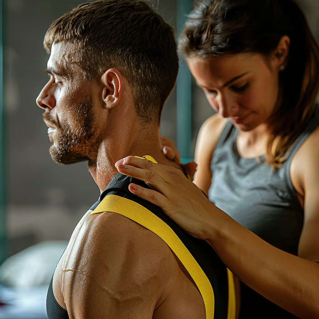
The evidence regarding Kinesio tape remains limited. A 2012 review found no clinically significant pain relief in the included studies. These studies focused primarily on individuals without injuries and on a few shoulder conditions; they do not cover all uses of Kinesio tape.24 The tape can be a complementary treatment worth trying based on your response, but it is not a substitute for exercises.
Further reading: Kamper SJ, Henschke N. Kinesio taping for sports injuries. 2013.25
When patellofemoral pain worsens, temporarily reduce the activities that trigger it and continue with activities you can tolerate well. After a recent dislocation, first follow the instructions you received during your medical evaluation; resuming activities and exercises should come later, based on the range of motion you have been cleared for. A fixed 10% increase per week does not guarantee that you will avoid injury. Progression should take into account your usual activities and how your body responds; data on the link between the rate of progression and injuries remains inconclusive.26
For many people, targeted exercises relieve pain within a few weeks to a few months. The pace of improvement varies from person to person20. The program can evolve as you regain ease and confidence in your activities. If you’re experiencing knee pain, our physical therapists can assess your patellar biomechanics and develop a personalized treatment plan.
Patellofemoral pain may persist or recur for months or years, and prognostic studies commonly include follow-up periods of more than one year. Follow-up allows for reassessment of symptoms, activity limitations, and the treatment plan if difficultiespersist .²⁷, ²⁸
Need professional advice?
Our physical therapists can assess your symptoms and provide you with a personalized treatment plan.
Make an appointmentReferences
Links open in a new tab.
- Smith TO, Davies L, Donell ST. The reliability and validity of assessing medio-lateral patellar position: a systematic review. Man Ther. 2009;14(4):355-62. (Back to section: 1)
- Kaufer H. Mechanical function of the patella. J Bone Joint Surg Am. 1971;53(8):1551-60. (Back to section: 1)
- Dai C, Pan L, Cao Y, Wang Q, Hong S. Autologous distal femoral condyle graft for patellar reconstruction during TKA in posttraumatic ankylosis: a case report. Front Surg. 2026;13:1791207. (Back to section: 1)
- Escamilla RF, Fleisig GS, Zheng N, Barrentine SW, Wilk KE, Andrews JR. Biomechanics of the knee during closed kinetic chain and open kinetic chain exercises. Med Sci Sports Exerc. 1998;30(4):556-69. (Back to section: 1)
- Hart HF, Patterson BE, Crossley KM, Culvenor AG, Khan MCM, King MG, et al. May the force be with you: understanding how patellofemoral joint reaction force compares across different activities and physical interventions—a systematic review and meta-analysis. Br J Sports Med. 2022;56(9):521-530. (Back to section: 1)
- Shepherd DET, Seedhom BB. Thickness of human articular cartilage in joints of the lower limb. Annals of the Rheumatic Diseases. 1999;58(1):27-34. (Back to section 1)
- Duong V, Oo WM, Ding C, Culvenor AG, Hunter DJ. Evaluation and Treatment of Knee Pain. JAMA. 2023;330(16):1568. (Back to section: 1)
- Rabelo NDDA, Lucareli PRG. Do hip muscle weakness and dynamic knee valgus matter for the clinical evaluation and decision-making process in patients with patellofemoral pain? Braz J Phys Ther. 2018;22(2):105-109. (Back to sections: 1, 2)
- Smith TO, Bowyer D, Dixon J, Stephenson R, Chester R, Donell ST. Can the vastus medialis oblique be preferentially activated? A systematic review of electromyographic studies. Physiother Theory Pract. 2009;25(2):69-98. (Back to sections: 1, 2)
- Neal BS, Lack SD, Lankhorst NE, Raye A, Morrissey D, van Middelkoop M. Risk factors for patellofemoral pain: a systematic review and meta-analysis. Br J Sports Med. 2019;53(5):270-281. (Back to section: 1)
- Hikawa K, Shimizu R, Takahashi S. Features of Elite Athletes' High and Asymmetric Quadriceps Angles: A Cross-Sectional Study. Health Sci Rep. 2025;8(6):e70909. (Back to section: 1)
- Smith TO, Hunt NJ, Donell ST. The reliability and validity of the Q-angle: a systematic review. Knee Surgery, Sports Traumatology, Arthroscopy. 2008;16(12):1068-1079. (Back to section: 1)
- Wills AK, Ramasamy A, Ewins DJ, Etherington J. The incidence and occupational outcome of overuse anterior knee pain during army recruit training. J R Army Med Corps. 2004;150(4):264-9. (Back to section: 1)
- Abdo N, Youssef EF, Ibrahim MM. Efficacy of adding manual therapy to hip and knee exercises in patients with patellofemoral pain syndrome: a double-blind randomized controlled clinical trial. Scientific Reports. 2025;15(1):32655. (Back to section: 1)
- Rebolledo BJ, Nam D, Cross MB, Green DW, Sculco TP. Familial association of femoral trochlear dysplasia with recurrent bilateral patellar dislocation. Orthopedics. 2012;35(4):e574-9. (Back to section: 1)
- Felson DT. The sources of pain in knee osteoarthritis. Current Opinion in Rheumatology. 2005;17(5):624-628. (Back to sections: 1, 2)
- International Association for the Study of Pain. Terminology. (Back to section: 1)
- Morgan M, Nazemian V, Harrington K, Ivanusic JJ. Mini review: The role of sensory innervation of subchondral bone in osteoarthritis pain. Frontiers in Endocrinology. 2022;13:1047943. (Back to section: 1)
- Shellock FG, Mink JH, Deutsch AL, Fox JM. Patellar tracking abnormalities: clinical experience with kinematic MR imaging in 130 patients. Radiology. 1989;172(3):799-804. (Back to section: 1)
- van der Heijden RA, Lankhorst NE, van Linschoten R, Bierma-Zeinstra SM, van Middelkoop M. Exercise for treating patellofemoral pain syndrome. Cochrane Database of Systematic Reviews. 2015;2015(1). (Back to sections: 1, 2)
- Collins NJ, Barton CJ, van Middelkoop M, Callaghan MJ, Rathleff MS, Vicenzino BT, et al. 2018 Consensus Statement on Exercise Therapy and Physical Interventions (Orthoses, Taping, and Manual Therapy) to Treat Patellofemoral Pain: Recommendations from the 5th International Patellofemoral Pain Research Retreat, Gold Coast, Australia, 2017. Br J Sports Med. 2018;52(18):1170-1178. (Back to section: 1)
- Kaya D, Doral MN, Callaghan M. How can we strengthen the quadriceps femoris in patients with patellofemoral pain syndrome? Muscles Ligaments Tendons J. 2012;2(1):25-32. (Back to section: 1)
- Bockrath K, Wooden C, Worrell T, Ingersoll CD, Farr J. Effects of patella taping on patella position and perceived pain. Med Sci Sports Exerc. 1993;25(9):989-92. (Back to section: 1)
- Williams S, Whatman C, Hume PA, Sheerin K. Kinesio Taping in the Treatment and Prevention of Sports Injuries. Sports Medicine. 2012;42(2):153-164. (Back to section: 1)
- Kamper SJ, Henschke N. Kinesio taping for sports injuries. Br J Sports Med. 2013;47(17):1128-9. (Back to section: 1)
- Fredette A, Roy JS, Perreault K, Dupuis F, Napier C, Esculier JF. The Association Between Running Injuries and Training Parameters: A Systematic Review. J Athl Train. 2022;57(7):650-671. (Back to section: 1)
- Willy RW, Hoglund LT, Barton CJ, Bolgla LA, Scalzitti DA, Logerstedt DS, et al. Patellofemoral Pain. J Orthop Sports Phys Ther. 2019;49(9):CPG1-CPG95. (Back to section: 1)
- Neal BS, Bolgla LA, Lack SD, Bazett-Jones DM, Boling M, Cowan SM, et al. Prognosis of Patellofemoral Pain: A Systematic Review With Evidence- and Gap-Map. Journal of Orthopaedic & Sports Physical Therapy. 2025;55(10):661-670. (Back to section: 1)
Customer satisfaction is our top priority
At Physioactif, excellence guides everything we do, but our patients are the best ones to tell you about it. Take a look at their verified reviews to get a real sense of their experience.






Discover our physical therapy clinics
We have locations in several areas to better serve you.
Blainville
190 Bas-de-Sainte-Thérèse Road, Suite 110,
Blainville, Quebec
J7B 1A7
Located in Blainville, near Rosemère, the Physioactif clinic is easily accessible to residents of the area and the surrounding communities
Boucherville
690 Rue de Montbrun, Suite S,
Boucherville, Quebec
J4B 8H2
Located in Boucherville, the Physioactif clinic is easily accessible to people in the area
Laval
3224 Jean-Béraud Ave., Suite 220, Laval,
QC H7T 2S4
Located in Chomedey, in the heart of Laval, the Physioactif clinic is easily accessible to people in the area
Montreal
8801 Lajeunesse Street,
Montreal,
QC H2M 1R8
Located in Ahuntsic, near Villeray, the Physioactif clinic is easily accessible to residents of both neighborhoods
Saint-Eustache
180 25th Avenue, Suite
201 Saint-Eustache
QC J7P 2V2
Located in Saint-Eustache, the Physioactif clinic is easily accessible to residents of the area and the surrounding communities
Vaudreuil
21 Cité-des-Jeunes Boulevard, Suite 240,
Vaudreuil-Dorion, Quebec
J7V 0N3
Located in Vaudreuil-Dorion, the Physioactif clinic is easily accessible to people in the area
Make an appointment now