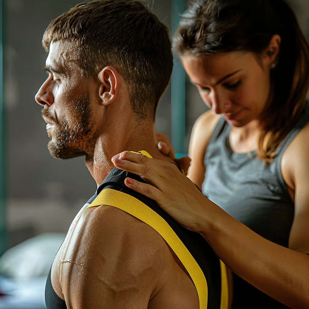
Patellofemoral syndrome
There is no clear definition of patellofemoral syndrome. It occurs when there is an overload between the femur and the patella due to a compression effect, which leads to irritation of this joint.
.png)
.png)
.png)



Patellofemoral syndrome, also known as “runner’s knee” or patellar syndrome, is one of the most common reasons for seeking medical attention for knee pain in sports medicine clinics1, accounting for 25 to 40 percent of all knee disorders2. Our half-squat arch control exercise targets a common contributing factor to this syndrome. This condition can affect both active and less active individuals. Our physical therapists support people with sports injuries as they return to their activities.
Here's the good news: with the right treatment, it's possible to resume your normal activities. Let's take a look at what might be causing this pain and how to relieve it.
What is Patellofemoral Pain Syndrome?
Patellofemoral syndrome is pain located around or behind the kneecap that is aggravated by activities that put stress on the knee. There is no single confirmed cause, and several factors may contribute to the symptoms.
10 Quick Tips to Understand Your Pain
One tip a day for 10 days to help you better understand your pain. About a 2-minute read.
The patella, the triangular-shaped bone located at the front of the knee, plays an essential role in movement. It acts as a pulley for the quadriceps, the muscle at the front of the thigh: it pulls the tendon away from the knee’s center of rotation, which increases its leverage for extending the knee3.
When this patella slips out of its groove, it is called dislocation or subluxation of the patella, an injury distinct from patellofemoral syndrome.What is the role of the kneecap in the knee?
The patella is a bone known as a “sesamoid” because it is located within the quadriceps tendon. This unique position allows it to facilitate the gliding motion of the tendon during movements in which you bend and straighten your knee.
The joint between the patella and the femur, the thigh bone, can withstand heavy loads.4 Pain may occur when activities exceed what the knee can handle at that moment, but several factors can contribute to it.
What causes Patellofemoral Pain Syndrome?
Patellofemoral syndrome can develop when activities that place stress on the knee increase or change. A rapid increase in training volume may contribute to it, but the evaluation takes several factors into account and does not assume a single cause.
In clinical practice, it is generally believed that increasing one’s running load too quickly increases the risk of overuse injuries, and training progression is adjusted based on this principle. A review of 31 studies published in 2012 was unable to determine which training characteristics were linked to injuries, as the results were contradictory5. An increase in training intensity may contribute to symptoms, but it does not explain all cases of pain. In 2025, a follow-up study linked a marked increase in the distance of a single run to more running injuries. It does not prove a single cause of patellofemoral syndrome.6 Another follow-up study, conducted among 629 runners, found that a lack of prior running experience was, in both men and women, the most significant risk factor for injury, without directly measuring the rate of increase in training load7.
The knee may become more sensitive after an increase in activity. Adjusting the level of exertion and allowing time for recovery can help. Progressive exercises can also improve mobility; complete rest is generally not the goal of treatment.
This strain can occur in several ways. A significant increase in mileage, an unusually long downhill run, or resuming vigorous exercise after a break can increase the stress placed on the knee. Your training progression should take into account your fitness level and your symptoms.
| Change in Business Activity | Adjust the tension as needed |
|---|---|
| Mountain hiking without training | A long descent can put unusual strain on the knee |
| Start of running season | A readjustment with a significant distance may exceed the current tolerance |
| New sport with jumping | Basketball, volleyball, soccer: gradually resume jumping and sprinting |
What are the risk factors for Patellofemoral Pain Syndrome?
Weakness in the quadriceps—the muscle that extends the knee—has been linked to a higher risk of patellofemoral syndrome in some studies, particularly among military recruits. However, the risk factors remain poorly understood.
Patellofemoral syndrome is common among active adolescents and young adults.8 Studies do not support the idea that this pain is caused by the femur growing faster than the surrounding tissues.
Jumping and sprinting can trigger pain in a person who is already experiencing symptoms. Weakness in the hip muscles—particularly those that abduct the leg—may be observed during an evaluation. This finding does not prove that it caused the problem.
Weakness in the quadriceps has been linked to an increased risk in follow-up studies, but this finding alone cannot predict who will develop this pain.9
What are the symptoms of Patellofemoral Pain Syndrome?
Pain in the front of the knee is the main symptom of patellofemoral syndrome. It usually develops gradually and worsens when you squat, go up or down stairs, run, or sit for long periods of time.
The pain is often diffuse and difficult to pinpoint precisely, a characteristic that the leading clinical practice guideline describes as pain that often begins gradually and is difficult to locate, situated in front of or around thepatella.¹⁰, ¹¹ Patients typically describe it as being “around” or “behind” the patella rather than at a specific point. The pain often returns after sitting for a long time—at the movies or in a car, for example—though the time it takes for the pain to appear varies greatly from person to person.
| Activity | Possible symptoms |
|---|---|
| Deep Squat | May increase pain when the knee is bent while bearing weight |
| Going down stairs | May cause pain in the front of the knee |
| Climbing stairs | May cause pain in the front of the knee |
| Running | Repeated pressure can worsen symptoms |
| Prolonged sitting | Keeping your knee bent may trigger pain |
How is Patellofemoral Pain Syndrome diagnosed?
The diagnosis of patellofemoral syndrome is based on the patient’s history of symptoms and a clinical examination performed by a healthcare professional. An X-ray is generally not necessary to confirm this diagnosis, but it may be indicated if the evaluation suggests another condition.1
The clinical evaluation includes a detailed review of your symptom history: when the pain began, what makes it worse, and what makes it better. Your physical therapist will observe the movements that trigger your symptoms and may check for tenderness around the kneecap. Kneecap compression tests are of limited use; no single test can confirm this diagnosis.
People whose imaging results are completely normal may still experience knee pain. A normal imaging result does not rule out pain.2
Magnetic resonance imaging, or MRI, produces detailed images of the inside of the knee. It is rarely necessary for this syndrome, but it can be helpful when another problem is suspected, such as a tear in a meniscus—a cartilage pad—or a ligament—a band that connects two bones. To learn more about another common knee condition, see our guide on patellar tendinopathy.
How does physiotherapy treat Patellofemoral Pain Syndrome?
Physical therapy treats patellofemoral syndrome primarily through progressive exercises and guidance on adjusting your activities. Your physical therapist will assess your range of motion, movement patterns, and strength to tailor the program to your needs. Mobilization techniques, exercises guided by the physical therapist, or coordination exercises may be added as needed.
Exercises form the basis of treatment, and their effectiveness is supported by research.12 In a trial involving 54 sedentary women aged 20 to 40, adding hip exercises to the knee exercise program further reduced pain and improved function13. Results of non-surgical treatment vary. In a five- to eight-year follow-up, 34 of the 60 respondents—57%—rated their recovery as poor. Only 19% of the initial group had responded14, and in another study of girls who had experienced anterior knee pain during adolescence, about one-quarter still reported significant symptoms 14 to 20years later15, 2.
| Assessed element | Assessment objective |
|---|---|
| Joint mobility | Identify movement restrictions |
| Nerve-related signs, if symptoms warrant it | Check for loss of sensation or strength and determine whether further evaluation is necessary |
| Movement Quality | Pay attention to how you squat and walk |
| Strength and stability | Assess knee and hip muscles |
Personalized treatment may include joint mobilization to improve the gliding motion of the patella, strengthening exercises that target the quadriceps and gluteal muscles, and exercises to improve your coordination. Education on how to manage the intensity of your activities is also an integral part of the treatment plan.
Patient education is an important part of treatment. It focuses on managing the physical load and pacing activities.10
Why is hip strengthening so important?
Strengthening the hip muscles can help treat patellofemoral syndrome. The gluteal muscles—particularly the gluteus medius—control the position of the femur during movement. One hypothesis suggests that a change in hip control alters the distribution of forces at the knee, but this mechanism has not been established as a causal chain16. What is better supported is the combination of hip and knee exercises to improve pain and function.
The program can combine knee and hip exercises based on your needs. The benefits of these exercises do not prove that the pain is caused by misalignment. A gradual and structured progression allows you to increase the intensity based on your response.
Does physical therapy work for patellofemoral syndrome?
Physiotherapy is recognized as an effective treatment for patellofemoral pain syndrome, with results supported by scientific research.
Physical therapy involves exercises that target both the hip and the knee to reduce pain.10 Exercises and activity recommendations form the foundation of treatment. Manual therapy may be added to this program depending on your symptoms and preferences.
The 2018 consensus recommends exercise—particularly exercises that involve both the hip and knee—to reduce pain and improve function. It also recommends certain combinations of interventions tailored to the individual12. The 2019 practice guideline supports knee and hip exercises10.
Responses to treatment vary from person to person. An assessment allows us to tailor the exercises to the patient’s symptoms, abilities, and desired activities, and then adjust them based on measured progress.
What exercises help relieve Patellofemoral Pain Syndrome?
Exercises that strengthen the hip and knee can reduce pain and improve function. Cycling or swimming can also help you stay physically active, depending on your tolerance and how your symptoms respond.
Knee and hip exercises can strengthen your muscles and make it easier for you to perform daily activities. A progressive exercise program tailors the movements and intensity to your specific situation.
| Strategy | Benefit |
|---|---|
| Glute strengthening | Strengthening the hip muscles; may help reduce pain |
| Swimming or cycling, depending on your tolerance | Continue physical activity if symptoms allow; adjust duration and intensity |
| Temporary knee brace | May provide relief for some people; its effect on the progression of the syndrome remains uncertain12 |
| Temporarily adjust activities that worsen symptoms | Temporarily adjust the load on the knee |
Your progress depends on your tolerance. The program may include muscle contractions without moving the knee, exercises where you bend and straighten the knee, and then movements specific to your sport. Your physical therapist will adjust the range of motion, resistance, and frequency based on your symptoms and progress.
How long does recovery take?
Recovery time depends on several factors: the severity of your symptoms, your activity level, and how consistently you follow the exercise program. Improvement is often seen after a few weeks of physical therapy, but the timeline varies from person to person.
Some people experience relief within the first few weeks, but the timeframe varies. Consistency in exercise and adjusting activities are part of the follow-up care.
A decrease in pain helps guide your recovery, but exercise tolerance is also important. Your physical therapist can help you gradually increase your running or sports activity and adjust your program if your symptoms worsen.
Can you treat patellofemoral syndrome on your own?
Limitations of self-management:Self-management can help with mild patellofemoral syndrome by adjusting activities and starting exercises that are tolerable. An evaluation is recommended if pain persists, function decreases, or the diagnosis remains unclear.
Role of the physiotherapist:The physical therapist will evaluate your knee, your strength, and the movements that are important for your activities. The evaluation is designed to identify any signs that would warrant a follow-up consultation and to develop a progressive program tailored to your situation.
Combining follow-up care with at-home exercises:Follow-up care may combine a professional evaluation with a home exercise program that is adjusted as progress is made.
When should you see a physiotherapist for patellofemoral pain syndrome?
See a physical therapist if your pain persists or limits your activities despite reasonable accommodations. In private practice in Quebec, you can see a physical therapist directly without a doctor’s referral; however, certain administrative or insurance requirements may call for a referral. Seek medical evaluation promptly if your knee is very painful, severely swollen, or deformed; if you cannot move it or put weight on it; if it locks or gives way; or if you have a fever or chills along with a red or warm knee.
Direct access to physical therapy allows you to see a physical therapist in a private clinic without a prior doctor's referral. If your condition requires further medical evaluation, your physical therapist will be able to refer you to the appropriate healthcare professional.
Physical therapy is an effective treatment for this condition. However, a complete recovery is not guaranteed. For many people, pain may persist for several years after rehabilitation.2 An exercise program tailored to your needs and adjusted based on your response can support your recovery.
Need professional advice?
Our physical therapists can assess your condition and provide you with a personalized treatment plan.
Make an appointmentReferences
The references in this article include consensus statements, practice guidelines, and studies on patellofemoral syndrome.
Links open in a new tab.
- Crossley KM, Stefanik JJ, Selfe J, Collins NJ, Davis IS, Powers CM, et al. 2016 Consensus Statement on Patellofemoral Pain from the 4th International Patellofemoral Pain Research Retreat, Manchester. Part 1: Terminology, definitions, clinical examination, natural history, patellofemoral osteoarthritis, and patient-reported outcome measures. Br J Sports Med. 2016;50(14):839-43. (Back to sections: 1, 2)
- Kasitinon D, Li WX, Wang EXS, Fredericson M. Physical Examination and Patellofemoral Pain Syndrome: An Updated Review. Curr Rev Musculoskelet Med. 2021;14(6):406-412. (Back to sections: 1, 2, 3, 4)
- Grelsamer RP, Weinstein CH. Applied biomechanics of the patella. Clin Orthop Relat Res. 2001(389):9-14. (Back to section: 1)
- Reilly DT, Martens M. Experimental analysis of quadriceps muscle force and patellofemoral joint reaction force during various activities. Acta Orthop Scand. 1972;43(2):126-37. (Back to section: 1)
- Nielsen RO, Buist I, Sørensen H, Lind M, Rasmussen S. Training errors and running-related injuries: a systematic review. Int J Sports Phys Ther. 2012;7(1):58-75. (Back to section: 1)
- Schuster Brandt Frandsen J, Hulme A, Parner ET, Møller M, Lindman I, Abrahamson J, et al. How much running is too much? Identifying high-risk running sessions in a 5,200-person cohort study. Br J Sports Med. 2025;59(17):1203-1210. (Back to section: 1)
- Buist I, Bredeweg SW, Bessem B, van Mechelen W, Lemmink KA, Diercks RL. Incidence and risk factors of running-related injuries during preparation for a 4-mile recreational running event. Br J Sports Med. 2010;44(8):598-604. (Back to section 1)
- Witvrouw E, Lysens R, Bellemans J, Cambier D, Vanderstraeten G. Intrinsic risk factors for the development of anterior knee pain in an athletic population. A two-year prospective study. Am J Sports Med. 2000;28(4):480-9. (Back to section: 1)
- Pappas E, Wong-Tom WM. Prospective Predictors of Patellofemoral Pain Syndrome: A Systematic Review with Meta-analysis. Sports Health. 2012;4(2):115-20. (Back to section: 1)
- Willy RW, Hoglund LT, Barton CJ, Bolgla LA, Scalzitti DA, Logerstedt DS, et al. Patellofemoral Pain. J Orthop Sports Phys Ther. 2019;49(9):CPG1-CPG95. (Back to sections: 1, 2, 3, 4)
- Cook C, Mabry L, Reiman MP, Hegedus EJ. Best tests and clinical findings for screening and diagnosing patellofemoral pain syndrome: a systematic review. Physiotherapy. 2012;98(2):93-100. (Back to section: 1)
- Collins NJ, Barton CJ, van Middelkoop M, Callaghan MJ, Rathleff MS, Vicenzino BT, et al. 2018 Consensus Statement on Exercise Therapy and Physical Interventions (Orthoses, Taping, and Manual Therapy) to Treat Patellofemoral Pain: Recommendations from the 5th International Patellofemoral Pain Research Retreat, Gold Coast, Australia, 2017. Br J Sports Med. 2018;52(18):1170-1178. (Back to sections: 1, 2, 3)
- Fukuda TY, Melo WP, Zaffalon BM, Rossetto FM, Magalhães E, Bryk FF, et al. Strengthening of the posterolateral hip muscles in sedentary women with patellofemoral pain syndrome: a randomized controlled clinical trial with a 1-year follow-up. J Orthop Sports Phys Ther. 2012;42(10):823-30. (Back to section: 1)
- Lankhorst NE, van Middelkoop M, Crossley KM, Bierma-Zeinstra SM, Oei EH, Vicenzino B, et al. Factors that predict a poor outcome 5–8 years after the diagnosis of patellofemoral pain: a multicenter observational analysis. Br J Sports Med. 2016;50(14):881–6. (Back to section: 1)
- Nimon G, Murray D, Sandow M, Goodfellow J. Natural history of anterior knee pain: a 14- to 20-year follow-up of nonoperative management. J Pediatr Orthop. 1998;18(1):118-22. (Back to section: 1)
- Powers CM. The influence of abnormal hip mechanics on knee injury: a biomechanical perspective. J Orthop Sports Phys Ther. 2010;40(2):42-51. (Back to section: 1)
.jpg)
Other conditions

Hip osteoarthritis is the normal wear and tear of the hip joint. It is often said that osteoarthritis is the wear and tear of the cartilage between our bones. That is true, but it involves more than just the cartilage. Cartilage is a tissue that acts as a cushion between the surfaces of our bones and allows our joints to glide smoothly and move fluidly.
This is normal wear and tear of the knee joint. It’s often said that osteoarthritis is the wearing down of the cartilage between our bones. That’s true, but it’s more than just the cartilage. Cartilage is a tissue that acts as a cushion between the surfaces of our bones and allows our joints to glide smoothly and move fluidly.
It is an inflammation of the subacromial bursa in the shoulder joint.
A bursa is a small, thin sac filled with fluid that is found in many of the body's joints. This small sac acts as a cushion within the joint and lubricates the structures that are subject to increased friction.
It is a tissue that surrounds the shoulder and helps keep the shoulder bone in place within the joint. The capsule helps keep the joint stable.
Neck pain is a general term used to describe pain in the neck that has no specific cause, such as an accident or a sudden awkward movement. Neck pain is therefore synonymous with “my neck hurts, and nothing in particular happened.”
In both types of injury, pain is felt in the neck and then radiates into the arm, or vice versa.
Make an appointment now
We offer a three-pronged quality assurance approach: optimized treatment time, a second opinion from a physical therapist, and ongoing expertise to ensure effective care tailored to your needs.

Customer satisfaction is our top priority
At Physioactif, excellence guides everything we do, but our patients are the best ones to tell you about it. Take a look at their verified reviews to get a real sense of their experience.






Discover our physical therapy clinics
We have locations in several areas to better serve you.
Blainville
190 Bas-de-Sainte-Thérèse Road, Suite 110,
Blainville, Quebec
J7B 1A7
Located in Blainville, near Rosemère, the Physioactif clinic is easily accessible to residents of the area and the surrounding communities
Boucherville
690 Rue de Montbrun, Suite S,
Boucherville, Quebec
J4B 8H2
Located in Boucherville, the Physioactif clinic is easily accessible to people in the area
Laval
3224 Jean-Béraud Ave., Suite 220, Laval,
QC H7T 2S4
Located in Chomedey, in the heart of Laval, the Physioactif clinic is easily accessible to people in the area
Montreal
8801 Lajeunesse Street,
Montreal,
QC H2M 1R8
Located in Ahuntsic, near Villeray, the Physioactif clinic is easily accessible to residents of both neighborhoods
Saint-Eustache
180 25th Avenue, Suite
201 Saint-Eustache
QC J7P 2V2
Located in Saint-Eustache, the Physioactif clinic is easily accessible to residents of the area and the surrounding communities
Vaudreuil
21 Cité-des-Jeunes Boulevard, Suite 240,
Vaudreuil-Dorion, Quebec
J7V 0N3
Located in Vaudreuil-Dorion, the Physioactif clinic is easily accessible to people in the area
Make an appointment now