Lumbar osteoarthritis

You've just received an X-ray or MRI report. Words like "arthritis," "degenerative changes," or "wear and tear" might have worried you. They suggest permanent damage and can make you think the pain will only get worse. It's normal to be concerned when you read these terms on a medical report.
Here's the good news: osteoarthritis is a normal part of aging, like gray hair or wrinkles.¹ Studies show that 60% of adults aged 40 have osteoarthritis visible on their scans, but many experience no pain.⁹ Structural changes don't always match symptoms.²
What Research Shows:- People with severe osteoarthritis on their scans may not experience any pain at all.³
- Conversely, those with minimal changes might experience a lot of discomfort.³
- Function can improve significantly with physiotherapy, despite the changes visible on imaging.⁷⁴
This guide explains what lumbar osteoarthritis truly means, why imaging doesn't determine your future, and how physiotherapy helps you maintain your function and reduce pain.
What is Low Back Osteoarthritis?
Lumbar osteoarthritis is the normal wear and tear of cartilage in the facet joints and disc spaces of your lower back. It causes pain, stiffness, and reduced mobility. This normal degenerative process affects most adults over 60. However, it can be managed very well with the right treatment and activity modifications.
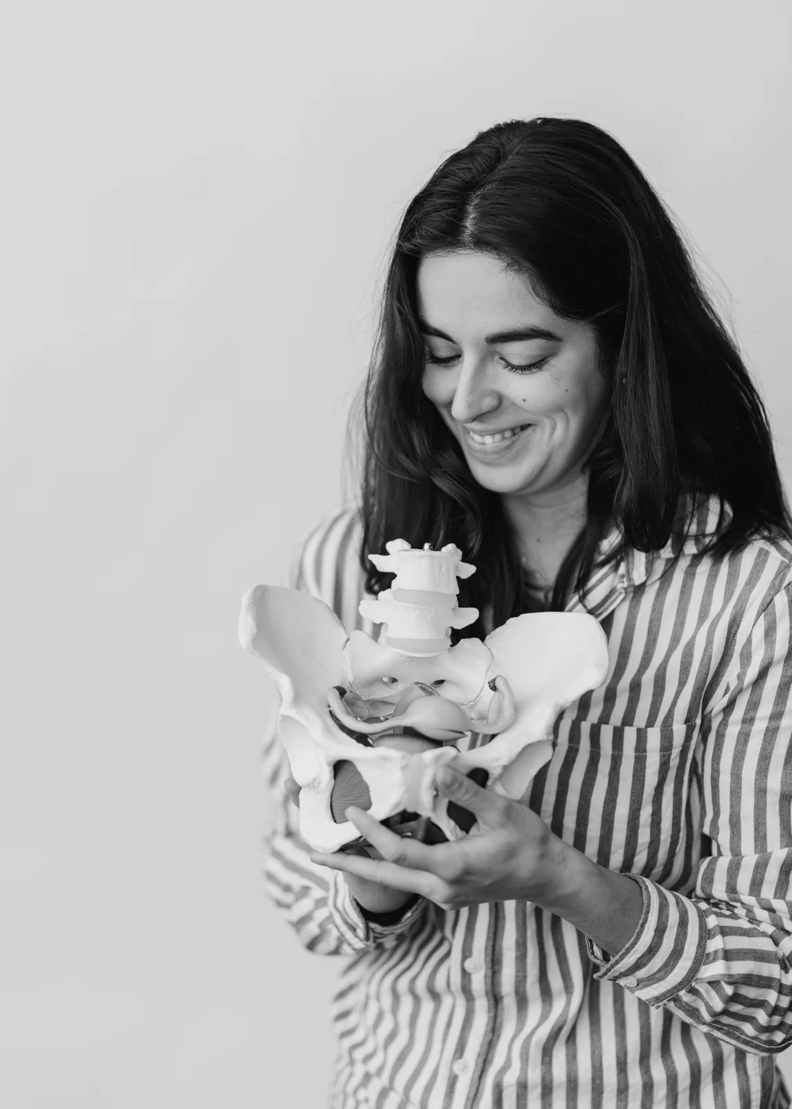
Osteoarthritis develops when cartilage wears down over time. Cartilage is the smooth tissue that covers joint surfaces. In the lumbar spine, it primarily affects the facet joints.⁴ These are small pairs of joints located at the back of each vertebra. They control how your spine moves.
Facet joints contain fluid and cartilage, making them similar to your knee or hip joints.⁵ As cartilage wears away, bones can develop small growths called osteophytes (bone spurs). The joint space also narrows. These changes appear on medical images and are often referred to as "degenerative disc disease" or "spondylosis" when they also affect the discs between your vertebrae.⁶
Osteoarthritis vs. Inflammatory Arthritis
Lumbar osteoarthritis is a wear-and-tear condition, not an inflammatory arthritis like rheumatoid arthritis.⁷ Degenerative arthritis develops due to repeated stress and aging. Inflammatory arthritis occurs when your immune system attacks your joints.⁸ This distinction is important because treatment approaches vary significantly. Our education about your condition helps you demystify osteoarthritis and adopt the right behaviors.
Studies show that lumbar osteoarthritis is extremely common. Imaging reveals facet joint osteoarthritis in over 60% of adults aged 40-49. Almost all adults over 80 have it.⁹ Yet, many of these individuals report no back pain. This clearly shows that structural changes are not the same as symptoms.¹⁰ To understand your lower back pain, you need to look beyond the images.
What Causes the Development of Osteoarthritis in the Lower Back?
Low back osteoarthritis develops due to repeated mechanical stress, aging, genetics, past injuries, and lifestyle factors. All these elements affect the cartilage in your joints. The cartilage degrades, bones remodel, and inflammation occurs. This leads to structural changes that vary greatly from person to person. Importantly, these changes have a very weak link to the level of pain you actually feel.
Daily Mechanical Stress
Your lower back supports heavy loads during daily activities. This is especially true at the L4-L5 and L5-S1 levels, where most movement occurs.¹¹ Repeated loading over decades slowly wears down cartilage surfaces, triggering bone changes. Jobs involving heavy lifting, prolonged sitting, or vibrations can accelerate this process. However, the relationship is complex and not entirely predictable.¹²
The Role of Genetics
Genetics plays a significant role in the risk of osteoarthritis. Twin studies suggest that about 50 to 65% of the risk of osteoarthritis is inherited. It affects the composition of your cartilage. It influences your bone density. It changes how you respond to inflammation.¹³ If your parents developed osteoarthritis, you are more likely to develop it too. However, this does not determine whether you will experience pain.
The Impact of Past Injuries
Past injuries can initiate or accelerate arthritic changes. Vertebral fractures, disc herniations, or ligament sprains alter how joints move and bear weight. This can trigger earlier cartilage degradation in affected areas.¹⁴ However, many people with significant injury histories never develop painful osteoarthritis. Others without any injury history still develop it.
Factors You Can Control
You can control certain risk factors: obesity, lack of movement, and smoking.
- Body Weight: Extra weight increases pressure on the facet joints, accelerating cartilage wear.¹⁵
- Movement: Surprisingly, complete rest also promotes osteoarthritis. It reduces cartilage nutrition. Cartilage needs movement to stay healthy.¹⁶
- Smoking: Smoking slows tissue healing and can accelerate degeneration due to its effects on blood vessels.¹⁷
Disc degeneration and facet joint osteoarthritis usually develop together. As discs lose height and water with age, the facet joints experience altered loading.¹⁸ This link means that "disc disease" and "facet osteoarthritis" often occur simultaneously.
What are the symptoms of lumbar osteoarthritis?
Symptoms include morning stiffness lasting less than 30 minutes, a dull back pain that worsens with activity, reduced flexibility, and sometimes grinding sensations during movement. If you recognize these symptoms, rest assured: they are very common and respond well to active treatment. Pain usually remains in the lower back without major leg symptoms, unless stenosis develops.
Typical Morning Stiffness
Morning stiffness is typical of osteoarthritis. After long periods of rest, your joints feel tight. Moving seems difficult. However, symptoms usually improve within 15 to 30 minutes once you start moving.¹⁹ This differs from inflammatory arthritis, where stiffness lasts an hour or more. The stiffness results from a temporary thickening of joint fluid and reduced joint lubrication during rest.²⁰
Osteoarthritis vs. Inflammatory Arthritis: How to Differentiate Them
| Characteristic | Osteoarthritis (degenerative) | Inflammatory arthritis |
|---|---|---|
| Morning Stiffness | Less than 30 minutes | More than an hour |
| Cause | Wear and aging | Immune system |
| Progression | Slow, progressive | Variable, in flare-ups |
| Improvement | With light movement | With initial rest |
Pain Patterns Throughout the Day
Pain patterns often follow mechanical patterns. Discomfort increases with long periods of standing, walking, or bending backward. These movements put stress on the facet joints.²¹ Sitting can initially provide relief (as it unloads the facet joints), but sitting for too long often causes discomfort. The pain is usually described as deep, dull, or stiff, rather than sharp or burning.²² This pattern of pain worsened by extension is explained in detail in our guide on directional preferences.
Pure Osteoarthritis vs. Nerve Compression
Unlike nerve compression issues, pure osteoarthritis rarely causes major symptoms in the legs. The pain usually stays in the lower back. It may spread to the buttocks or upper thighs, but it doesn't follow specific nerve pathways.²³ However, when bone spurs or thickened ligaments press on the spinal nerve passages, spinal stenosis can develop.
Functional Limitations
Functional limitations develop slowly. You might notice a reduced ability to bend forward or backward. You might also have difficulty getting up from a chair. These limitations are often more related to pain avoidance and muscle guarding than to actual joint damage.²⁴ This means they can significantly improve with the right treatment, even if structural changes persist.
Key Point : The severity of symptoms does not match the severity of osteoarthritis seen on images. Research consistently shows a weak link between X-ray findings and pain levels.²⁵10 Quick Tips for Understanding Your Pain
The ones that have made the biggest difference in my patients' lives. 1 a day, 2 minutes.
How is lumbar osteoarthritis diagnosed?
Diagnosis combines a clinical examination, which reveals reduced mobility and local tenderness, with X-ray results. However, imaging results don't always match symptoms. This makes clinical evaluation essential for planning treatment. We cannot rely solely on structural findings.
Your physiotherapist or doctor looks for several typical signs during the examination. They check your spinal mobility and note any reduced range of motion when you bend forward, backward, or rotate.²⁷ They palpate to find tenderness over the facet joints, which are located approximately 2 to 3 cm from the center of your lower back.²⁸ Movements that involve bending backward usually cause pain, while bending forward can provide relief.²⁹ Our movement re-education approach specifically targets these restrictions.
What changes on X-rays indicate lumbar osteoarthritis?
X-rays show the structural signs of osteoarthritis:
- Narrowing of the joint space as cartilage thins
- Formation of osteophytes (bone spurs) at the edges of the joints
- Subchondral sclerosis (increased bone density beneath the cartilage)
- Subchondral cysts (fluid-filled sacs within the bone)
Radiologists often classify the severity of osteoarthritis from mild to severe based on these findings.³⁰ CT scans provide more detailed views of bone changes. MRI shows cartilage, joint inflammation, and soft tissue problems.³¹
Why don't imaging results always match symptoms?
The mismatch between imaging and symptoms is one of the most important concepts for managing lumbar osteoarthritis. Large population studies show that people without symptoms often have the same imaging results as those with severe pain.³² A major study found facet joint osteoarthritis in 89% of adults over 60. Yet, many reported no back pain.³³
This mismatch occurs because pain depends on several factors beyond just structure:
- Muscle Conditioning
- Movement Patterns
- Nervous System Sensitivity
- Psychological Factors
- Sleep Quality
- Stress Levels³⁴
How does physiotherapy treat osteoarthritis and how does exercise help?
Physiotherapy Treatments:Physiotherapy combines manual therapy, targeted exercises, and education to manage symptoms. Treatment focuses on keeping you functional rather than reversing osteoarthritis.
Joint mobilizations and manipulations improve the mobility of facet joints.³⁶ Soft tissue work reduces muscle tension.³⁷
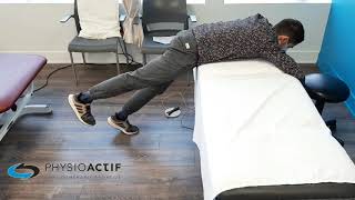
Therapeutic exercise forms the foundation of management. Muscle strengthening and endurance exercises strengthen the core and hip muscles that support your spine.³⁸ Flexibility exercises maintain your range of motion.³⁹ Aerobic exercise promotes cartilage health.⁴⁰
Education about pain science empowers you to take control. Our personalized education program demystifies chronic pain.⁴¹ A multimodal approach consistently shows better results than a single treatment.⁴³
Exercise as the most effective treatment:Exercise is the most effective treatment for osteoarthritis. It reduces pain and improves function despite joint changes. Cartilage gets its nutrients during joint compression and decompression.⁴⁵ Regular exercise keeps cartilage healthier than rest.⁴⁶ Appropriate loading protects cartilage.⁴⁷
Several types of exercise benefit osteoarthritis: stabilizing muscle exercises for the core,⁴⁸ flexibility exercises,⁴⁹ and aerobic activities. The McKenzie approach offers a structured method of directional exercises.
Dosage matters: keep discomfort below 3-4 out of 10 during exercise.⁵¹ Progression should be slow.⁵³ A little discomfort is normal.⁵⁴ The benefits last as long as you continue exercising.⁵⁷
Book an appointment for a comprehensive assessment and a personalized treatment plan.What lifestyle changes help manage lumbar osteoarthritis?
Key changes include maintaining a healthy weight to reduce joint load, staying physically active with low-impact activities, improving your posture and workspace setup, using heat for stiffness, and pacing your activities to avoid flare-ups.
Weight Management
Weight management profoundly affects osteoarthritis symptoms. Every extra kilogram of body weight increases pressure on the lumbar joints, multiplying that pressure during movement.⁵⁸ A weight loss of just 5 to 10% of body weight can significantly reduce pain and improve function.⁵⁹
Activity Choices
Choosing and modifying activities helps you stay active without worsening your symptoms. Low-impact activities like swimming, aqua fitness, cycling, or using an elliptical machine provide cardiovascular benefits while reducing stress on your joints.⁶⁰ A complete postural analysis can reveal imbalances that worsen your osteoarthritis.
Workspace Setup
Setting up your workspace properly reduces continuous stress during daily activities. A well-adjusted desk and chair can help reduce long periods spent in one position. Alternate between sitting and standing throughout the day.⁶² Our ergonomic workstation assessment service identifies specific adjustments for your professional situation.
Using Heat
Heat works well to reduce stiffness and improve movement. Morning showers, heating pads, or warm pools help prepare tissues for activity.⁶⁴ Heat is usually more beneficial before activity.
Pacing Activities
Pacing yourself and conserving energy can prevent symptom flare-ups. Alternate between more strenuous and easier activities. Take regular movement breaks. Increase activity levels slowly.⁶⁶ Pacing doesn't mean avoiding activity; it means distributing activity intelligently.
Need professional advice?
Our physical therapists can assess your condition and provide you with a personalized treatment plan.
Make an appointmentHow does lumbar osteoarthritis progress over time?
Osteoarthritis typically progresses slowly over years, with periods of stability and occasional flare-ups. While structural changes continue, symptoms don't always worsen at the same rate. Many individuals maintain good function through active management. Symptoms often stabilize rather than constantly deteriorating.
Studies on the natural history of lumbar osteoarthritis show that its progression varies greatly and is often unpredictable.⁶⁷ Some people show rapid progression on X-rays with minimal changes in symptoms. Others have stable imaging results but changing symptoms.⁶⁸ This indicates that structural progression and symptom progression are, in part, separate processes.
Flare-up management
Flare-ups (periods of increased pain and stiffness) sometimes occur in most people with osteoarthritis. This is normal and does not mean your condition is worsening. They are often triggered by new activities, poor sleep, or stress.⁶⁹ Strategies include temporary changes in activities (not complete rest), increased use of heat or ice, and continuing gentle movement.⁷¹
Factors Affecting Progression
Ongoing obesity, smoking, complete lack of activity, and poor muscle conditioning can accelerate symptoms.⁷² On the other hand, maintaining a healthy weight, staying physically active, and managing other conditions like diabetes or depression can slow down symptom progression.⁷³
The outlook for maintaining function is generally good with active management. Long-term studies show that most people with lumbar osteoarthritis maintain their functional abilities over 5 to 10 years, especially when they engage in regular exercise and physiotherapy.⁷⁴When to consider other treatments and how to distinguish from facet joint syndrome?
Other treatment options:Consider other treatments when physiotherapy alone doesn't adequately control symptoms after 8-12 weeks. Facet injections offer temporary relief.⁷⁶ Radiofrequency ablation provides longer relief for some patients.⁷⁷ NSAIDs are effective short-term but carry gastrointestinal risks.⁸⁰ Surgery remains a last resort.⁸¹
If your osteoarthritis results from a work accident, CNESST covers 100% of treatments. Our telerehabilitation service offers complete remote sessions.
Distinction from facet joint syndrome:Osteoarthritis involves chronic degenerative changes visible on scans. Facet joint syndrome describes acute irritation, often without structural changes.⁸⁶ Osteoarthritis develops slowly over months or years. Facet joint syndrome often occurs suddenly. Many patients have both conditions simultaneously.
How to regain control over your lumbar osteoarthritis?
Our physiotherapists create personalized, evidence-based programs to reduce pain, improve mobility, and keep you active. The osteoarthritis visible on your scans will remain, but symptoms can significantly improve through physiotherapy, exercise, and lifestyle changes.
We help you move from fear of diagnosis to confident self-management. If your condition stems from a car accident, our services are covered by SAAQ.
Book an appointment to schedule your assessment and create a personalized program.References
- Brinjikji W, Luetmer PH, Comstock B, et al. Systematic literature review of imaging features of spinal degeneration in asymptomatic populations. AJNR Am J Neuroradiol. 2015;36(4):811-816.
- Kalichman L, Li L, Kim DH, et al. Facet joint osteoarthritis and low back pain in the community-based population. Spine. 2008;33(23):2560-2565.
- Suri P, Miyakoshi A, Hunter DJ, et al. Does lumbar spinal degeneration begin with the anterior structures? BMC Musculoskelet Disord. 2011;12:202.
- Bogduk N. Clinical and Radiological Anatomy of the Lumbar Spine. 5th ed. Edinburgh: Churchill Livingstone; 2012.
- Jaumard NV, Welch WC, Winkelstein BA. Spinal facet joint biomechanics and mechanotransduction. J Biomech Eng. 2011;133(7):071010.
- Kirkaldy-Willis WH, Wedge JH, Yong-Hing K, Reilly J. Pathology and pathogenesis of lumbar spondylosis and stenosis. Spine. 1978;3(4):319-328.
- Goldring MB, Goldring SR. Osteoarthritis. J Cell Physiol. 2007;213(3):626-634.
- Felson DT. Osteoarthritis as a disease of mechanics. Osteoarthritis Cartilage. 2013;21(1):10-15.
- Suri P, Miyakoshi A, Hunter DJ, et al. Does lumbar spinal degeneration begin with the anterior structures? BMC Musculoskelet Disord. 2011;12:202.
- Kalichman L, Li L, Kim DH, et al. Facet joint osteoarthritis and low back pain in the community-based population. Spine. 2008;33(23):2560-2565.
- Nachemson AL. Disc pressure measurements. Spine. 1981;6(1):93-97.
- Bakker EW, Verhagen AP, Lucas C, et al. Daily spinal mechanical loading as a risk factor for acute non-specific low back pain. Eur Spine J. 2007;16(1):107-113.
- Battié MC, Videman T, Levälahti E, et al. Genetic and environmental effects on disc degeneration. Spine. 2008;33(25):2801-2808.
- Adams MA, Roughley PJ. What is intervertebral disc degeneration, and what causes it? Spine. 2006;31(18):2151-2161.
- Shiri R, Karppinen J, Leino-Arjas P, et al. The association between obesity and low back pain: a meta-analysis. Am J Epidemiol. 2010;171(2):135-154.
- Holm S, Maroudas A, Urban JP, et al. Nutrition of the intervertebral disc. Connect Tissue Res. 1981;8(2):101-119.
- Behrend C, Prasarn M, Coyne E, et al. Smoking cessation related to improved patient-reported pain scores. J Bone Joint Surg Am. 2012;94(23):2161-2166.
- Fujiwara A, Lim TH, An HS, et al. The effect of disc degeneration and facet joint osteoarthritis on segmental flexibility. Spine. 2000;25(23):3036-3044.
- Hootman JM, Helmick CG. Projections of US prevalence of arthritis and associated activity limitations. Arthritis Rheum. 2006;54(1):226-229.
- Buckwalter JA, Saltzman C, Brown T. The impact of osteoarthritis: implications for research. Clin Orthop Relat Res. 2004;(427 Suppl):S6-15.
- Manchikanti L, Boswell MV, Singh V, et al. Prevalence of facet joint pain in chronic spinal pain. BMC Musculoskelet Disord. 2004;5:15.
- Schwarzer AC, Aprill CN, Derby R, et al. Clinical features of patients with pain stemming from the lumbar zygapophysial joints. Spine. 1994;19(10):1132-1137.
- Bogduk N. International Spinal Injection Society guidelines for spinal injection procedures. Clin J Pain. 1997;13(4):285-302.
- Gellhorn AC, Katz JN, Suri P. Osteoarthritis of the spine: the facet joints. Nat Rev Rheumatol. 2013;9(4):216-224.
- Kalichman L, Li L, Kim DH, et al. Facet joint osteoarthritis and low back pain in the community-based population. Spine. 2008;33(23):2560-2565.
- O'Sullivan P. It's time for change with the management of non-specific chronic low back pain. Br J Sports Med. 2012;46(4):224-227.
- Cook C, Hegedus E, Showalter C, Sizer PS Jr. Coupling behavior of the lumbar spine. J Manipulative Physiol Ther. 2006;29(7):554-558.
- Dreyfuss P, Michaelsen M, Pauza K, et al. The value of medical history and physical examination in diagnosing sacroiliac joint pain. Spine. 1996;21(22):2594-2602.
- Laslett M, Öberg B, Aprill CN, McDonald B. Zygapophysial joint blocks in chronic low back pain. BMC Musculoskelet Disord. 2004;5:43.
- Kellgren JH, Lawrence JS. Radiological assessment of osteo-arthrosis. Ann Rheum Dis. 1957;16(4):494-502.
- Weishaupt D, Zanetti M, Hodler J, Boos N. MR imaging of the lumbar spine. Radiology. 1998;209(3):661-666.
- Brinjikji W, Luetmer PH, Comstock B, et al. Systematic literature review of imaging features of spinal degeneration. AJNR Am J Neuroradiol. 2015;36(4):811-816.
- Kalichman L, Li L, Kim DH, et al. Facet joint osteoarthritis and low back pain. Spine. 2008;33(23):2560-2565.
- Moseley GL, Butler DS. Fifteen years of explaining pain. J Pain. 2015;16(9):807-813.
- Fairbank JC, Pynsent PB. The Oswestry Disability Index. Spine. 2000;25(22):2940-2952.
- Bronfort G, Haas M, Evans R, et al. Effectiveness of manual therapies. Chiropr Osteopat. 2010;18:3.
- Bialosky JE, Bishop MD, Price DD, et al. The mechanisms of manual therapy. Man Ther. 2009;14(5):531-538.
- Searle A, Spink M, Ho A, Chuter V. Exercise interventions for the treatment of chronic low back pain. Clin Rehabil. 2015;29(12):1155-1167.
- Hayden JA, van Tulder MW, Tomlinson G. Systematic review: strategies for using exercise therapy. Ann Intern Med. 2005;142(9):776-785.
- Dunn KM, Jordan KP, Croft PR. Contributions of prognostic factors for poor outcome. Eur J Pain. 2011;15(3):313-319.
- Louw A, Diener I, Butler DS, Puentedura EJ. The effect of neuroscience education on pain. Arch Phys Med Rehabil. 2011;92(12):2041-2056.
- Vlaeyen JW, Linton SJ. Fear-avoidance and its consequences. Pain. 2000;85(3):317-332.
- van Middelkoop M, Rubinstein SM, Verhagen AP, et al. Exercise therapy for chronic nonspecific low-back pain. Best Pract Res Clin Rheumatol. 2010;24(2):193-204.
- Foster NE, Anema JR, Cherkin D, et al. Prevention and treatment of low back pain. Lancet. 2018;391(10137):2368-2383.
- Urban JP. The role of the physicochemical environment in determining disc cell behaviour. Biochem Soc Trans. 2002;30(Pt 6):858-864.
- Belavý DL, Albracht K, Bruggemann GP, et al. Can exercise positively influence the intervertebral disc? Sports Med. 2016;46(4):473-485.
- Holm S, Maroudas A, Urban JP, et al. Nutrition of the intervertebral disc. Connect Tissue Res. 1981;8(2):101-119.
- Hides JA, Richardson CA, Jull GA. Multifidus muscle recovery. Spine. 1996;21(23):2763-2769.
- Marshall PW, Kennedy S, Brooks C, Lonsdale C. Pilates exercise or stationary cycling for chronic nonspecific low back pain. Spine. 2013;38(15):E952-959.
- Hurwitz EL, Morgenstern H, Chiao C. Effects of recreational physical activity. Am J Public Health. 2005;95(10):1817-1824.
- Smith BE, Hendrick P, Smith TO, et al. Should exercises be painful in the management of chronic musculoskeletal pain? Br J Sports Med. 2017;51(23):1679-1687.
- American College of Sports Medicine. ACSM's Guidelines for Exercise Testing and Prescription. 10th ed. Philadelphia: Wolters Kluwer; 2018.
- Nijs J, Roussel N, van Wilgen CP, et al. Thinking beyond muscles and joints. Man Ther. 2013;18(2):96-102.
- Smith BE, Hendrick P, Smith TO, et al. Should exercises be painful? Br J Sports Med. 2017;51(23):1679-1687.
- Henchoz Y, Kai-Lik So A. Exercise and nonspecific low back pain. Joint Bone Spine. 2008;75(5):533-539.
- Jordan JL, Holden MA, Mason EE, Foster NE. Interventions to improve adherence to exercise. Cochrane Database Syst Rev. 2010;(1):CD005956.
- Bassett SF. The assessment of patient adherence to physiotherapy rehabilitation. N Z J Physiother. 2003;31(2):60-66.
- Shiri R, Karppinen J, Leino-Arjas P, et al. The association between obesity and low back pain. Am J Epidemiol. 2010;171(2):135-154.
- Christensen R, Bartels EM, Astrup A, Bliddal H. Effect of weight reduction in obese patients. Ann Rheum Dis. 2007;66(4):433-439.
- Dundar U, Solak O, Yigit I, et al. Clinical effectiveness of aquatic exercise. Spine. 2009;34(14):1436-1440.
- Hurwitz EL, Morgenstern H, Chiao C. Effects of recreational physical activity. Am J Public Health. 2005;95(10):1817-1824.
- Robertson M, Amick BC 3rd, DeRango K, et al. The effects of an office ergonomics training. Appl Ergon. 2009;40(1):124-135.
- Claus AP, Hides JA, Moseley GL, Hodges PW. Different ways to balance the spine. Spine. 2009;34(6):E208-214.
- French SD, Cameron M, Walker BF, et al. Superficial heat or cold for low back pain. Cochrane Database Syst Rev. 2006;(1):CD004750.
- Nadler SF, Weingand K, Kruse RJ. The physiologic basis of cryotherapy and thermotherapy. Pain Physician. 2004;7(3):395-399.
- Murphy SL, Smith DM, Clauw DJ, Alexander NB. The impact of momentary pain and fatigue. Arthritis Rheum. 2008;59(6):849-856.
- Suri P, Hunter DJ, Rainville J, et al. Presence and extent of severe facet joint osteoarthritis. Osteoarthritis Cartilage. 2013;21(9):1199-1206.
- Fujiwara A, Lim TH, An HS, et al. The effect of disc degeneration and facet joint osteoarthritis. Spine. 2000;25(23):3036-3044.
- Hawker GA, Stewart L, French MR, et al. Understanding the pain experience. Osteoarthritis Cartilage. 2008;16(4):415-422.
- Parry E, Ogollah R, Peat G. Significant pain variability in persons with knee osteoarthritis. BMC Musculoskelet Disord. 2017;18(1):80.
- Bannuru RR, Osani MC, Vaysbrot EE, et al. OARSI guidelines for non-surgical management. Osteoarthritis Cartilage. 2019;27(11):1578-1589.
- Sandmark H, Nisell R. Validity of five common manual neck pain provoking tests. Scand J Rehabil Med. 1995;27(3):131-136.
- Foster NE, Anema JR, Cherkin D, et al. Prevention and treatment of low back pain. Lancet. 2018;391(10137):2368-2383.
- Hartvigsen J, Hancock MJ, Kongsted A, et al. What low back pain is and why we need to pay attention. Lancet. 2018;391(10137):2356-2367.
- Foster NE, Anema JR, Cherkin D, et al. Prevention and treatment of low back pain. Lancet. 2018;391(10137):2368-2383.
- Manchikanti L, Pampati V, Fellows B, Bakhit CE. The diagnostic validity of facet joint nerve blocks. Curr Rev Pain. 2000;4(5):337-344.
- Maas ET, Ostelo RW, Niemisto L, et al. Radiofrequency denervation for chronic low back pain. Cochrane Database Syst Rev. 2015;(10):CD008572.
- American College of Rheumatology. Recommendations for medical management of osteoarthritis. Arthritis Rheum. 2000;43(9):1905-1915.
- Zhang W, Moskowitz RW, Nuki G, et al. OARSI recommendations for management of osteoarthritis. Osteoarthritis Cartilage. 2008;16(2):137-162.
- Chou R, McDonagh MS, Nakamoto E, Griffin J. Analgesics for Osteoarthritis. Rockville: AHRQ; 2011.
- Mirza SK, Deyo RA. Systematic review of randomized trials comparing lumbar fusion surgery. Spine. 2007;32(7):816-823.
- Jacobs W, Van der Gaag NA, Tuschel A, et al. Total disc replacement for chronic back pain. Cochrane Database Syst Rev. 2012;(9):CD008326.
- Deyo RA, Mirza SK, Martin BI, et al. Trends, complications, and charges for lumbar spinal stenosis surgery. JAMA. 2010;303(13):1259-1265.
- Vickers AJ, Vertosick EA, Lewith G, et al. Acupuncture for chronic pain. J Pain. 2018;19(5):455-474.
- Astin JA. Why patients use alternative medicine. JAMA. 1998;279(19):1548-1553.
- Manchikanti L, Boswell MV, Singh V, et al. Prevalence of facet joint pain. BMC Musculoskelet Disord. 2004;5:15.
- Schwarzer AC, Aprill CN, Derby R, et al. Clinical features of lumbar facet syndrome. Spine. 1994;19(10):1132-1137.
- Schwarzer AC, Wang SC, Bogduk N, et al. Prevalence and clinical features of lumbar facet joint pain. Ann Rheum Dis. 1995;54(2):100-106.
- Gellhorn AC, Katz JN, Suri P. Osteoarthritis of the spine: the facet joints. Nat Rev Rheumatol. 2013;9(4):216-224.
- Friedrich KM, Nemec S, Peloschek P, et al. The prevalence of lumbar facet joint edema. Skeletal Radiol. 2007;36(8):755-760.
- Kalichman L, Li L, Kim DH, et al. Facet joint osteoarthritis and low back pain. Spine. 2008;33(23):2560-2565.
- Cohen SP, Raja SN. Pathogenesis, diagnosis, and treatment of lumbar facet joint pain. Anesthesiology. 2007;106(3):591-614.
- Gellhorn AC, Katz JN, Suri P. Osteoarthritis of the spine: the facet joints. Nat Rev Rheumatol. 2013;9(4):216-224.
- Bronfort G, Haas M, Evans R, et al. Effectiveness of manual therapies. Chiropr Osteopat. 2010;18:3.


Other conditions
Hip osteoarthritis is the normal wear and tear of the hip joint. It is often said that osteoarthritis is the wear and tear of the cartilage between our bones. That is true, but it involves more than just the cartilage. Cartilage is a tissue that acts as a cushion between the surfaces of our bones and allows our joints to glide smoothly and move fluidly.
This is normal wear and tear of the knee joint. It’s often said that osteoarthritis is the wearing down of the cartilage between our bones. That’s true, but it’s more than just the cartilage. Cartilage is a tissue that acts as a cushion between the surfaces of our bones and allows our joints to glide smoothly and move fluidly.
It is an inflammation of the subacromial bursa in the shoulder joint.
A bursa is a small, thin sac filled with fluid that is found in many of the body's joints. This small sac acts as a cushion within the joint and lubricates the structures that are subject to increased friction.
It is a tissue that surrounds the shoulder and helps keep the shoulder bone in place within the joint. The capsule helps keep the joint stable.
Neck pain is a general term used to describe pain in the neck that has no specific cause, such as an accident or a sudden awkward movement. Neck pain is therefore synonymous with “my neck hurts, and nothing in particular happened.”
In both types of injury, pain is felt in the neck and then radiates into the arm, or vice versa.
It is a severe strain or tear of the muscle fibers in the groin or inner thigh.
Make an appointment now
We offer a three-pronged quality assurance approach: optimized treatment time, a second opinion from a physical therapist, and ongoing expertise to ensure effective care tailored to your needs.

Customer satisfaction is our top priority
At Physioactif, excellence guides everything we do, but our patients are the best ones to tell you about it. Take a look at their verified reviews to get a real sense of their experience.
Discover our physical therapy clinics
We have locations in several areas to better serve you.
Blainville
190 Bas-de-Sainte-Thérèse Road, Suite 110,
Blainville, Quebec
J7B 1A7
Located in Blainville, near Rosemère, the Physioactif clinic is easily accessible to residents of the area and the surrounding communities
Laval
3224 Jean-Béraud Ave., Suite 220, Laval,
QC H7T 2S4
Located in Chomedey, in the heart of Laval, the Physioactif clinic is easily accessible to people in the area
Montreal
8801 Lajeunesse Street,
Montreal,
QC H2M 1R8
Located in Ahuntsic, near Villeray, the Physioactif clinic is easily accessible to residents of both neighborhoods
Saint-Eustache
180 25th Avenue, Suite
201 Saint-Eustache
QC J7P 2V2
Located in Saint-Eustache, the Physioactif clinic is easily accessible to residents of the area and the surrounding communities
Vaudreuil
21 Cité-des-Jeunes Boulevard, Suite 240,
Vaudreuil-Dorion, Quebec
J7V 0N3
Located in Vaudreuil-Dorion, the Physioactif clinic is easily accessible to people in the area
Make an appointment now
