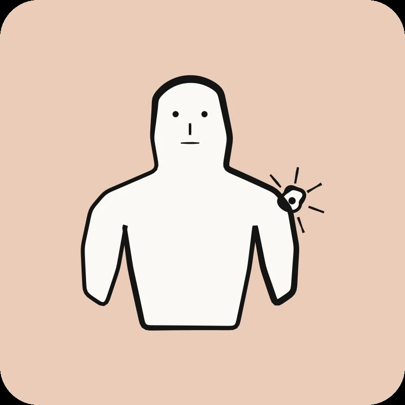
Shoulder Bursitis: Treatment and Recovery in Physio
What is shoulder bursitis?
Shoulder bursitis is an inflammation of the subacromial bursa, a small fluid-filled sac located between the acromion (the upper bony part of the shoulder blade) and the rotator cuff tendons. This bursa acts as a protective cushion, facilitating the smooth movement of structures when lifting the arm.
When this bursa becomes irritated or inflamed, it thickens and produces excess fluid, which reduces the available space in the shoulder joint. This inflammation causes the characteristic pain of bursitis, especially when lifting the arm or at night.
There are two main types of shoulder bursitis:
- Acute bursitis: Sudden inflammation, often caused by direct trauma (e.g., falling on the shoulder) or intensive overuse (e.g., painting a ceiling for several hours). The pain is intense and appears quickly.
- Chronic bursitis: Persistent inflammation that develops gradually over several weeks or months, usually due to repetitive movements or an untreated subacromial impingement.
It is important to distinguish bursitis from shoulder tendinitis, although the two conditions often coexist. Bursitis affects the bursa (a protective structure), while tendinitis directly impacts the rotator cuff tendons. Symptoms can be similar, but treatment may vary slightly depending on the primary structure affected.
In reality, about 60 to 70% of patients diagnosed with bursitis also have rotator cuff involvement7, which is why a comprehensive evaluation by a physiotherapist is essential to identify all involved structures.
Now that you understand what bursitis is, let's look at the causes that can lead to this inflammation.
What are the causes of shoulder bursitis?
Shoulder bursitis is caused by repetitive overhead arm movements, direct trauma, subacromial impingement, inflammatory arthritis, or sometimes an infection. Professional and sports activities involving repeated motions significantly increase the risk.
Repetitive Movements: The Most Common Cause
The main cause of shoulder bursitis is repeated stress on the joint, especially during arm movements above 90 degrees of elevation. Each time you lift your arm overhead, the subacromial space narrows, which can compress the bursa.
At-risk activities and professions include:
- Sports: Swimming (especially freestyle), baseball, tennis, volleyball, throwing sports
- Professions: Painting, construction (overhead work), hairdressing, carpentry, electrical work
- Daily Activities: Prolonged gardening, window washing, frequently storing items overhead
When these movements are repeated without adequate recovery periods, the bursa gradually becomes irritated, thickens, and develops chronic inflammation.
Subacromial Impingement: The Primary Mechanism
Subacromial impingement is a reduction of the space between the acromion and the head of the humerus (upper arm bone). This reduced space compresses the bursa when the arm is raised, creating repeated friction that leads to inflammation.
This impingement can be caused by:
- Poor posture (shoulders rounded forward)
- Weakness in the rotator cuff muscles
- Stiffness in the posterior joint capsule
- A particular anatomical shape of the acromion (some people have a more hooked acromion)
- Shoulder blade (scapula) instability
Subacromial impingement is present in about 75% of chronic bursitis cases8, making it the most important biomechanical mechanism to address in physiotherapy.
Direct Trauma
A fall onto the shoulder or a direct impact can cause acute traumatic bursitis. The impact suddenly compresses the bursa, triggering an immediate inflammatory reaction. This type of bursitis typically presents with intense pain, visible swelling, and a sudden limitation of movement.
Inflammatory arthritis
Certain systemic conditions increase the risk of developing bursitis:
- Gout: Deposits of uric acid crystals in the bursa
- Rheumatoid Arthritis: Chronic inflammation affecting joints and bursae
- Psoriatic Arthritis: Inflammation associated with psoriasis
In these cases, bursitis is part of a broader clinical picture requiring joint medical management.
Infection (Septic Bursitis): Rare but Serious
Although rare (less than 5% of bursitis cases9), infectious (or septic) bursitis can occur after a penetrating wound, shoulder surgery, or in individuals with weakened immune systems. It presents with intense pain, fever, local redness, and warmth to the touch. This condition requires urgent medical treatment with antibiotics.
Risk Factors to Consider
Certain factors increase the likelihood of developing bursitis:
- Age: The highest incidence occurs between 40 and 60 years old, due to the cumulative wear and tear on the structures.
- Occupation: Jobs involving repetitive overhead arm movements increase the risk by 3 to 4 times10.
- High-Intensity Sports: Competitive swimmers and throwers have bursitis rates 5 to 6 times higher than the general population11.
Understanding these causes helps you better identify what might have triggered your bursitis. Let's now look at how this inflammation typically presents in daily life.
What are the symptoms of shoulder bursitis?
Symptoms of shoulder bursitis include throbbing pain at rest and at night, morning joint stiffness, local swelling, painful limitation of overhead movements, and difficulty sleeping on the affected side.
Characteristic Pain: Throbbing and Nighttime
The pain associated with bursitis has distinct characteristics that set it apart from other shoulder conditions:
- Quality: Dull, throbbing pain, deep pressure sensation (rather than sharp, stabbing pain)
- Location: Outer side of the shoulder, sometimes radiating up the arm, but rarely beyond the elbow
- Timing: Significantly worse at night and at rest, which disrupts sleep
Nighttime pain is particularly characteristic of bursitis. It is explained by an increase in pressure within the bursa when the arm is still, combined with increased inflammation during the night. Many patients report that they cannot sleep on the affected side and wake up multiple times during the night.
The Painful Arc: A Key Diagnostic Sign
A very telling symptom of bursitis is the 'painful arc'. This refers to a specific range of arm elevation (typically between 60 and 120 degrees of lateral abduction) where the pain is at its worst.
Here's what happens: between 60 and 120 degrees, the inflamed bursa is maximally compressed between the acromion and the head of the humerus. Below 60 degrees and above 120 degrees, the compression is less, so the pain decreases. This painful arc phenomenon helps your physiotherapist differentiate bursitis from other shoulder conditions.
Joint Stiffness, Especially in the Morning
Shoulder stiffness is particularly noticeable upon waking and generally improves with gentle movement throughout the day. This stiffness primarily affects:
- Lateral arm elevation (abduction)
- External rotation (such as reaching behind your head)
- Internal rotation (such as fastening a bra or reaching for a back pocket)
Unlike adhesive capsulitis (frozen shoulder), bursitis stiffness is mainly due to pain and muscle guarding, rather than an actual loss of joint range of motion. With treatment, this stiffness usually resolves within a few weeks.
Swelling and Loss of Strength
Although less common, visible or palpable swelling can appear in the acute phase, especially in cases of traumatic or infectious bursitis.
Many patients also report a loss of strength, but it's important to understand that this weakness is generally secondary to pain (pain inhibition) rather than actual muscle damage. When pain decreases with treatment, strength typically returns without intensive training.
Recognizing these symptoms helps you better understand what you're experiencing. But how do we confirm it's actually bursitis?
10 Quick Tips for Understanding Your Pain
The ones that have made the biggest difference in my patients' lives. 1 a day, 2 minutes.
How is shoulder bursitis diagnosed?
Diagnosing shoulder bursitis relies on a clinical examination by a physiotherapist or doctor, which includes specific tests (Neer, Hawkins-Kennedy), palpation of the painful area, and sometimes imaging (ultrasound, MRI) to confirm inflammation.
Clinical Examination: The Cornerstone of Diagnosis
Your physiotherapist begins with a comprehensive assessment that includes:
- Detailed Case History: Onset of symptoms, triggering factors, professional and sports activities, history of injuries
- Postural Observation: Shoulder position at rest, asymmetries, overall posture
- Palpation: Identifying precise painful areas, detecting swelling or local heat
- Range of Motion: Assessing active movements (which you perform yourself) and passive movements (which the therapist guides)
- Muscle Strength Tests: To evaluate rotator cuff function
Specific Tests for Bursitis
Several clinical tests help identify bursitis:
Neer Test: The therapist passively raises your arm in internal rotation. If this reproduces your pain, the test suggests subacromial impingement with bursitis.
Hawkins-Kennedy Test: The arm is placed at 90 degrees of forward flexion, then the therapist performs a forced internal rotation. This movement compresses the bursa and causes pain if it is inflamed.
Painful Arc: The therapist observes your active lateral elevation movement to identify the area of maximum pain (typically 60-120 degrees).
These tests have a combined sensitivity of 80-85% for detecting subacromial pathologies, including bursitis12.
Differential Diagnosis: Distinguishing Bursitis from Other Conditions
It is essential to differentiate bursitis from other shoulder conditions that present similar symptoms:
- Rotator Cuff Tendinitis: Pain during specific rotational movements, more pronounced muscle weakness
- Subacromial Impingement: A mechanism often causing bursitis, but can exist without bursa inflammation
- Adhesive Capsulitis (Frozen Shoulder): Significant loss of passive range of motion, progression through three distinct phases
- Glenohumeral Osteoarthritis: Progressive pain, crepitus (cracking sounds), limitation of all movements
The good news is that in 60-70% of cases, bursitis coexists with tendinitis or impingement13, meaning that physiotherapy treatment addressing all these elements is often necessary and effective.
Imaging: When Is It Necessary?
In most cases, a clinical diagnosis is sufficient to begin treatment. However, imaging can be useful in certain situations:
Musculoskeletal Ultrasound: This is the preferred examination for visualizing the bursa. It allows us to see wall thickening, the presence of fluid, and the condition of the rotator cuff. Ultrasound is quick, cost-effective, and radiation-free.
MRI: Recommended if the diagnosis is uncertain, if symptoms do not improve after 4 to 6 weeks of treatment, or if a rotator cuff tear is suspected. MRI offers a detailed view of all deep structures of the shoulder.
X-ray: Although it does not show soft tissues like the bursa, X-rays are useful for ruling out bone conditions (osteoarthritis, calcifications, fractures) that could cause similar symptoms.
Important Point: Studies show that up to 30% of people without shoulder pain exhibit signs of bursal inflammation on ultrasound14. This means that imaging should always be interpreted in conjunction with your clinical symptoms, not in isolation.
Role of the Physiotherapist in Diagnosis
In Quebec, physiotherapists are first-line professionals who can diagnose bursitis without a prior medical referral. They are trained to:
- Identify musculoskeletal conditions
- Differentiate between conditions requiring conservative treatment and those needing medical investigation
- Refer to a doctor if warning signs are present (infection, massive tear, no improvement)
Now that the diagnosis is clear, let's explore how physiotherapy can effectively treat your bursitis.
How Does Physiotherapy Treat Shoulder Bursitis?
Physiotherapy treats shoulder bursitis with a progressive four-phase approach: pain and inflammation control, mobility recovery, rotator cuff strengthening, and recurrence prevention.
As physiotherapists specializing in shoulder conditions at Physioactif, we adapt this protocol based on the phase of your bursitis and your specific functional goals. To learn more about our therapeutic approach and what you can expect, consult our guide on physiotherapy for shoulder bursitis. Here's how we proceed.
Phase 1: Pain and Inflammation Control (0-2 weeks)
The initial goal is to calm acute inflammation and reduce pain to allow for the start of mobilization.
Relative Rest: This does not mean complete immobilization, but rather avoiding movements that reproduce pain, especially raising the arm above 90 degrees and lifting heavy loads. You can continue your daily activities as long as they do not cause significant pain.
Cryotherapy (Ice): Applying ice for 15 to 20 minutes, 3 to 4 times a day (especially after any activity), helps reduce inflammation, decrease pain, and control swelling. Ice is particularly effective in the first 48 to 72 hours.
Therapeutic Modalities:
- Therapeutic Ultrasound: Sound waves that penetrate deeply to reduce inflammation and improve local circulation
- Low-Intensity Laser: Helps reduce pain and accelerate tissue healing
- TENS (Transcutaneous Electrical Nerve Stimulation): Controls pain by modulating nerve signals
Gentle Manual Techniques: Low-amplitude joint mobilizations, massage of peripheral muscles (neck, shoulder blade) to release compensatory tensions, and lymphatic drainage to reduce swelling.
Phase 2: Mobility Recovery (2-4 weeks)
Once pain is better controlled, the goal is to regain full, pain-free range of motion.
Progressive Mobilizations: Your physiotherapist guides your arm through passive movements (you relax, they move your arm) and active-assisted movements (you participate in the movement with their help). This progression helps increase range of motion without worsening inflammation.
Codman (Pendulum) Exercises: These exercises use gravity to decompress the subacromial space. You lean forward, let your arm hang, and perform small circles or swings. Recommended frequency: 3 to 5 times a day, 2 to 3 minutes per session.
Progressive Capsular Stretches: Gentle stretches for the posterior joint capsule (often tight) and pectoral muscles (which contribute to rounded shoulders). Hold for 30 seconds, 3 to 4 repetitions, twice a day.
A 2022 systematic review shows that early mobilization (starting from the first week) reduces the total recovery time by 30% to 40% compared to strict rest15.
Phase 3: Strengthening and Stabilization (4-8 weeks)
Strengthening is key to preventing recurrence. This phase begins when pain is well-controlled and you have recovered at least 80% of your range of motion.
Rotator Cuff Strengthening: These four muscles (supraspinatus, infraspinatus, teres minor, subscapularis) stabilize the head of the humerus and prevent subacromial impingement. Exercises progress from light resistance bands to moderate weights, with a particular focus on external rotation (often the weakest).
Scapular Stabilization: The lower trapezius, serratus anterior, and rhomboids are strengthened to optimize shoulder blade positioning. A stable shoulder blade improves the subacromial space and reduces stress on the bursa.
Functional Exercises: These simulate daily and sports-specific movements related to your activities. For example, a swimmer will work on crawl stroke movement patterns with progressive resistance, while a painter will practice controlled overhead movements.
Postural Correction: Addressing forward head posture, rounded shoulders, and excessive thoracic kyphosis (rounded upper back). These postural issues reduce the subacromial space and increase the risk of recurrence.
Phase 4: Recurrence Prevention and Return to Activities (ongoing)
The final phase aims to consolidate your gains and prevent the bursitis from returning.
Maintenance Program: A home exercise program 2 to 3 times a week, combining rotator cuff strengthening and stretches. This program should be maintained for at least 6 to 12 months post-recovery.
Work and Sport Ergonomics: Adjusting workstation height, organizing your space to avoid repetitive overhead movements, taking regular breaks during shoulder-intensive activities, and correcting sports technique if necessary.
Gradual Return to Activities: Increasing volume and intensity according to the 10% rule (do not increase by more than 10% per week). Listen to your body's signals: pain greater than 2/10 after activity or increased morning stiffness indicates that you should slow down your progression.
Our shoulder physiotherapy approach integrates these four phases in a personalized way, based on your initial condition, functional goals, and response to treatment.
Let's now look at the specific exercises you can do in each phase.
Need professional advice?
Our physical therapists can assess your condition and provide you with a personalized treatment plan.
Make an appointmentWhat exercises are good for shoulder bursitis?
Exercises for shoulder bursitis include Codman (pendulum) exercises in the acute phase, progressive capsular stretches, progressive rotator cuff strengthening with resistance bands, and postural corrections to reduce subacromial impingement.
IMPORTANT: The exercises presented below are general recommendations. Each case of bursitis is different and requires a personalized program. Consult your physiotherapist to get an exercise plan tailored to your healing phase and specific limitations.
Acute Phase: Codman (Pendulum) Exercises
These exercises are safe from the first few days and help maintain minimal mobility without worsening inflammation.
Technique:
- Lean forward, supporting yourself on a table with your healthy hand.
- Let the affected arm hang loosely without contracting your shoulder muscles.
- Perform small circles (20-30 cm diameter), then swing your arm back and forth and side to side.
- Allow gravity to create passive movement; do not actively force it.
Dosage: 2 to 3 minutes, 4 to 5 times per day
Caution: You should not feel any pain during this exercise. If you experience pain, reduce your range of motion.
Subacute Phase: Capsular Stretches
These stretches begin once acute pain subsides (typically after 1 to 2 weeks).
Posterior Capsule Stretch:
- Bring your affected arm across your body (as if giving a hug).
- With your opposite hand, gently pull your elbow towards your opposite shoulder.
- You should feel a stretch at the back of your shoulder.
Hold: 30 seconds, 3 to 4 repetitions, 2 times per day
Pectoral Stretch:
- Place your forearm against a doorframe, with your elbow bent at 90 degrees.
- Turn your body in the opposite direction until you feel a stretch in your chest.
- This stretch is crucial because tight pectoral muscles can cause the shoulder to roll forward, reducing the space under the acromion (shoulder blade).
Strengthening Phase: Rotator Cuff with Resistance Band
These exercises strengthen the muscles that stabilize the shoulder and help prevent impingement.
External Rotation with Resistance Band:
- Attach a resistance band to a doorknob at elbow height.
- Stand sideways, with your elbow tucked against your body and bent at 90 degrees.
- Perform an external rotation against the resistance of the band (as if opening a door).
- Control the movement as you return to the starting position.
Dosage: 2 to 3 sets of 10 to 15 repetitions, start with light resistance.
Internal Rotation:
- Maintain the same position, but turn to face the other way.
- Perform an internal rotation (bringing your hand towards your stomach).
- The external rotation to internal rotation ratio should be 3:2, as external rotators are generally weaker and more important for stability.
Controlled Abduction:
- Stand with an elastic band under the foot of the affected side.
- Raise your arm sideways from 0 to 90 degrees.
- Control the descent (the eccentric phase is important for strengthening).
Caution: Do not shrug your shoulder during exercises. Maintain good alignment.
Stabilization Phase: Postural Exercises
These exercises correct postural issues that contribute to the impingement.
Scapular Retraction:
- Stand or sit with arms at your sides.
- Bring your shoulder blades together towards your spine (as if squeezing a pencil between them).
- Hold for 5 to 10 seconds, 10 to 15 repetitions.
Frequency: 3 to 4 times a day, especially if you sit for long periods.
Thoracic Extension:
- Use a foam roller placed horizontally under your upper back.
- Perform small extensions by leaning backward.
- This exercise corrects thoracic kyphosis and improves shoulder blade positioning.
Studies show that adding scapular stabilization exercises to standard bursitis treatment improves outcomes by 25-30% at 12 weeks16.
Once these exercises are mastered, how long does it take for a full recovery?
How long does shoulder bursitis last?
Acute shoulder bursitis typically lasts 2 to 6 weeks with appropriate physiotherapy treatment. Chronic or untreated cases can persist for several months, but full recovery is possible with adequate treatment.
Recovery Timeline: Acute Phase
For acute bursitis treated early, the typical progression is as follows:
- Week 1-2: Pain and inflammation control. Significant reduction in pain at rest and improved sleep.
- Week 3-4: Mobility recovery. Increased range of motion, decreased morning stiffness, ability to perform basic daily activities.
- Week 5-8: Strengthening and functional return. Recovery of strength, gradual return to sports and professional activities.
After 6 weeks, approximately 70% to 80% of patients treated with physiotherapy report a significant improvement in their symptoms17.
Factors that influence healing time
Several elements can either speed up or slow down your recovery:
Initial Severity: A mild bursitis might resolve in 2 to 3 weeks, while a severe case with significant swelling and marked limitation could require 6 to 8 weeks.
Early Treatment: Patients who start physiotherapy within the first week of symptom onset recover 30% to 40% faster than those who wait 4 weeks or more18.
Adherence to Exercise Program: Patients who regularly perform their home exercises (at least 5 days a week) recover on average 2 to 3 weeks faster than those who are inconsistent.
Presence of Associated Conditions: Isolated bursitis heals faster than bursitis accompanied by rotator cuff tendinitis or rotator cuff syndrome. In these cases, the duration can extend to 8 to 12 weeks.
Continuous Aggravating Factors: If you continue to perform the repetitive movements that caused the bursitis (unmodified overhead work, intensive sport), healing can be delayed by 50% to 100%.
Chronic Bursitis: A Different Challenge
Bursitis is considered chronic when symptoms persist beyond 3 months. The most common causes of chronicity are:
- Lack of treatment in the first few weeks
- Unmodified aggravating factors (ergonomics, sports technique)
- Unaddressed structural subacromial impingement
- Persistent rotator cuff weakness
The prognosis for chronic bursitis remains good, but recovery generally requires 3 to 6 months of intensive physiotherapy treatment. The important thing is not to give up: with adequate treatment and modification of contributing factors, even chronic bursitis can resolve completely.
Signs of Positive Progression
Here are the indicators that your treatment is working:
- Reduction in nighttime pain (often the first sign of improvement)
- Improved range of motion, especially in the morning
- Decrease in the frequency and intensity of pain
- Increased ability to perform daily activities
- Fewer nighttime awakenings
When to be concerned?
Consult your physiotherapist or doctor if:
- No improvement is noted after 2 to 3 weeks of physiotherapy treatment
- Symptoms worsen despite treatment
- New symptoms appear (numbness, significant weakness)
- You develop a fever (possible infection)
Now that you know the typical recovery time, let's look at when it's necessary to consult a professional.
When to consult for shoulder bursitis?
Consult a physiotherapist if your shoulder pain lasts more than a week, if you experience significant limitation in daily movements, night pain that prevents sleep, or a loss of strength in your arm.
Signs requiring prompt consultation
Here are the situations that warrant a physiotherapy consultation within days of symptoms appearing:
Persistent pain for more than a week: If your shoulder pain lasts beyond 7 days despite relative rest and applying ice, a professional evaluation is recommended. Pain that lasts more than a week suggests inflammation requiring active intervention.
Significant functional limitation: If you have difficulty performing basic daily activities such as getting dressed, styling your hair, reaching for overhead objects, or lifting light loads (2-3 kg), it's time to seek consultation.
Night pain disrupting sleep: Pain that wakes you up at night or prevents you from sleeping on the affected side is a sign of significant inflammation requiring treatment.
Progressive loss of strength: If you notice increasing weakness in your arm or difficulty holding your arm up, an evaluation is necessary to differentiate between pain-induced inhibition and a possible rotator cuff injury.
Significant swelling: Visible swelling or a marked increase in the size of your shoulder may indicate severe acute bursitis or septic (infectious) bursitis requiring immediate attention.
Intensification of pain at rest: If the pain worsens even when you are not using your shoulder, this may indicate a progression of the inflammation.
Warning signs requiring urgent medical consultation
Certain symptoms indicate potentially serious conditions that require an emergency medical evaluation:
Sudden and intense pain after trauma: Violent and sudden pain after a fall or direct impact may indicate a complete rotator cuff tear or even an associated fracture.
Total inability to move the arm: The inability to initiate active arm movement may signal a massive tendon tear or a mechanical blockage requiring immediate investigation.
Fever accompanied by shoulder pain: The combination of fever (>38°C), intense pain, local redness, and warmth to the touch suggests septic bursitis (infection of the bursa) which requires urgent antibiotics.
Neurological symptoms: Numbness, tingling that extends down the arm to the hand, or sudden hand weakness may indicate nerve compression (axillary nerve or brachial plexus) requiring rapid evaluation.
Benefits of early consultation
Consulting early offers several significant benefits:
Faster recovery: Studies show that patients treated within the first week have a total recovery time reduced by 30 to 40% compared to those who wait 4 weeks or more19.
Prevention of chronicity: Untreated bursitis becomes chronic in 15 to 20% of cases20. Early treatment interrupts the inflammation-pain-stiffness-weakness cycle before it becomes permanently established.
Accurate diagnosis: Only a professional evaluation can differentiate bursitis from tendinitis, subacromial impingement, or adhesive capsulitis (frozen shoulder). Each of these conditions has a slightly different prognosis and treatment.
Personalized Program: A physiotherapist creates a treatment plan tailored to your healing phase, specific limitations, functional goals, and activity level.
At Physioactif, our comprehensive assessment helps identify the exact cause of your shoulder pain and establish an evidence-based treatment plan, tailored to your condition and functional goals. Our physiotherapists, specializing in shoulder conditions, use manual techniques, therapeutic modalities, and progressive exercise programs to help you regain a functional, pain-free shoulder.
Once you have recovered, how can you prevent a recurrence?
How to prevent recurrent shoulder bursitis?
Preventing recurrent shoulder bursitis involves maintaining rotator cuff strength, practicing good ergonomics at work and during sports, adequate warm-up, and a gradual progression of loads and repetitive movements.
Maintain a Continuous Strengthening Program
Maintaining the strength and endurance of the rotator cuff muscles is the cornerstone of prevention.
Minimum Frequency: 2 to 3 times per week, even after full recovery. Each session should include rotator cuff strengthening exercises (external/internal rotation) and scapular stabilization (retraction, depression).
Recommended Duration: Maintain the program for at least 6 to 12 months post-recovery, ideally permanently if you engage in high-risk activities (overhead sports, demanding profession).
Prospective studies show that patients who maintain a long-term exercise program have a recurrence rate of less than 10%, compared to 30% to 40% in those who stop exercising after initial recovery21.
Work Ergonomics: Essential Modifications
If your job involves repetitive shoulder movements, a few simple adjustments can make a big difference:
Work Surface Height: Adjust your workstation to avoid working with your arms raised above 90 degrees for prolonged periods. Use step stools or ladders to reach high objects instead of stretching.
Regular Breaks: Take a 2 to 3-minute break every 30 to 45 minutes during repetitive tasks. Use this time to perform gentle stretches and scapular retraction exercises.
Space Organization: Place frequently used objects at a height between your shoulder and waist, thus avoiding repeated overhead movements.
Sports Ergonomics: Technique and Progression
For athletes, technique and progression are crucial:
Swimming: Work on your crawl stroke technique with a coach to optimize body rotation (and not just shoulder movement). Include a specific 10 to 15-minute shoulder warm-up before each training session.
Tennis and Racket Sports: Improve your serving technique by using your legs and trunk rotation more, rather than relying solely on your shoulder. Increase your training volume by a maximum of 10% per week.
Baseball and Throwing Sports: Work on your throwing mechanics with a qualified coach, perform preventive rotator cuff strengthening, and adhere to the recommended pitching limits based on your age and skill level.
Gradual Activity Progression: The 10% Rule
Whether returning to sports or increasing work volume, the 10% rule is essential: never increase the volume or intensity of your activities by more than 10% per week.
Example: If you swim 2000 meters per week, do not increase to more than 2200 meters the following week.
Signs to Slow Down: Pain greater than 2/10 after activity, or increased morning stiffness the next day, indicates that you are progressing too quickly.
Specific Warm-up: Non-Negotiable
Before any activity that intensely uses the shoulder:
Warm-up Content: Gentle shoulder movements for 5 minutes (arm circles, Codman exercises), rotator cuff activation with a light resistance band (2 sets of 10-15 repetitions), dynamic stretches for the pectorals and posterior capsule.
Minimum Duration: 10 to 15 minutes before any intense shoulder activity.
Daily posture correction
Sitting Position: Avoid slouching and a forward head posture when working at the computer. Place your screen at eye level, use lumbar support, and perform scapular retraction exercises regularly.
Phone and Computer Use: Avoid holding the phone between your shoulder and ear. Use headphones or a speaker for prolonged calls.
Recognizing early signs of recurrence
Pay attention to these warning signs that indicate a recurrence is starting:
- Mild but persistent pain during specific movements
- Morning stiffness that reappears
- Pain during movements that were previously comfortable
- Slight decrease in perceived strength
Recommended Action: Immediately resume your Phase 1 and 2 exercises (ice, gentle movements, Codman). If the pain persists for more than 3 to 5 days, consult your physiotherapist for a re-evaluation.
Periodic physiotherapy follow-up
Recommended Frequency: A follow-up assessment 3 to 6 months after initial recovery is recommended, especially if you engage in high-risk activities.
Follow-up Objectives: Adjust your exercise program according to your evolving needs, correct any postural or movement compensations that may have developed, and update your prevention plan.
Prevention can also apply to other shoulder conditions. To learn more about preventive strategies, consult our guide on shoulder tendinitis, which shares several similar prevention principles.
References
- Luime JJ, Koes BW, Hendriksen IJ, et al. Prevalence and incidence of shoulder pain in the general population; a systematic review. Scand J Rheumatol. 2004;33(2):73-81.
- Steuri R, Sattelmayer M, Elsig S, et al. Effectiveness of conservative interventions including exercise, manual therapy and medical management in adults with shoulder impingement: a systematic review and meta-analysis of RCTs. Br J Sports Med. 2017;51(18):1340-1347.
- Hanratty CE, McVeigh JG, Kerr DP, et al. The effectiveness of physiotherapy exercises in subacromial impingement syndrome: a systematic review and meta-analysis. Semin Arthritis Rheum. 2012;42(3):297-316.
- Neer CS 2nd. Anterior acromioplasty for the chronic impingement syndrome in the shoulder: a preliminary report. J Bone Joint Surg Am. 1972;54(1):41-50.
- Girish G, Lobo LG, Jacobson JA, et al. Ultrasound of the shoulder: asymptomatic findings in men. AJR Am J Roentgenol. 2011;197(4):W713-9.
- Coombes BK, Bisset L, Vicenzino B. Efficacy and safety of corticosteroid injections and other injections for management of tendinopathy: a systematic review of randomised controlled trials. Lancet. 2010;376(9754):1751-1767.
- Papatheodorou A, Ellinas P, Takis F, et al. US of the shoulder: rotator cuff and non-rotator cuff disorders. Radiographics. 2006;26(1):e23.
- Bigliani LU, Levine WN. Subacromial impingement syndrome. J Bone Joint Surg Am. 1997;79(12):1854-1868.
- Broy SB, Schmid FR. A comparison of medical drainage (needle aspiration) and surgical drainage (bursectomy or resection) in the initial treatment of infected bursa. Clin Orthop Relat Res. 1986;(210):103-107.
- van der Windt DA, Thomas E, Pope DP, et al. Occupational risk factors for shoulder pain: a systematic review. Occup Environ Med. 2000;57(7):433-442.
- McMaster WC, Troup J. A survey of interfering shoulder pain in United States competitive swimmers. Am J Sports Med. 1993;21(1):67-70.
- Park HB, Yokota A, Gill HS, et al. Diagnostic accuracy of clinical tests for the different degrees of subacromial impingement syndrome. J Bone Joint Surg Am. 2005;87(7):1446-1455.
- Lewis JS, Green AS, Dekel S. The aetiology of subacromial impingement syndrome. Physiotherapy. 2001;87(9):458-469.
- Michener LA, Walsworth MK, Doukas WC, Murphy KP. Reliability and diagnostic accuracy of 5 physical examination tests and combination of tests for subacromial impingement. Arch Phys Med Rehabil. 2009;90(11):1898-1903.
- Page MJ, Green S, McBain B, et al. Manual therapy and exercise for rotator cuff disease. Cochrane Database Syst Rev. 2016;(6):CD012224.
- Struyf F, Nijs J, Mollekens S, et al. Scapular-focused treatment in patients with shoulder impingement syndrome: a randomized clinical trial. Clin Rheumatol. 2013;32(1):73-85.
- Kuhn JE. Exercise in the treatment of rotator cuff impingement: a systematic review and a synthesized evidence-based rehabilitation protocol. J Shoulder Elbow Surg. 2009;18(1):138-160.
- Gebremariam L, Hay EM, van der Sande R, et al. Subacromial impingement syndrome: effectiveness of physiotherapy and manual therapy. Br J Sports Med. 2014;48(16):1202-1208.
- Rhon DI, Boyles RB, Cleland JA. One-year outcome of subacromial corticosteroid injection compared with manual physical therapy for the management of the unilateral shoulder impingement syndrome: a pragmatic randomized trial. Ann Intern Med. 2014;161(3):161-169.
- Littlewood C, May S, Walters S. Epidemiology of rotator cuff tendinopathy: a systematic review. Shoulder Elbow. 2013;5(4):256-265.
- Ludewig PM, Borstad JD. Effects of a home exercise programme on shoulder pain and functional status in construction workers. Occup Environ Med. 2003;60(11):841-849.
Videos in this category
Other conditions
Hip osteoarthritis is the normal wear and tear of the hip joint. It is often said that osteoarthritis is the wear and tear of the cartilage between our bones. That is true, but it involves more than just the cartilage. Cartilage is a tissue that acts as a cushion between the surfaces of our bones and allows our joints to glide smoothly and move fluidly.
This is normal wear and tear of the knee joint. It’s often said that osteoarthritis is the wearing down of the cartilage between our bones. That’s true, but it’s more than just the cartilage. Cartilage is a tissue that acts as a cushion between the surfaces of our bones and allows our joints to glide smoothly and move fluidly.
It is an inflammation of the subacromial bursa in the shoulder joint.
A bursa is a small, thin sac filled with fluid that is found in many of the body's joints. This small sac acts as a cushion within the joint and lubricates the structures that are subject to increased friction.
It is a tissue that surrounds the shoulder and helps keep the shoulder bone in place within the joint. The capsule helps keep the joint stable.
Neck pain is a general term used to describe pain in the neck that has no specific cause, such as an accident or a sudden awkward movement. Neck pain is therefore synonymous with “my neck hurts, and nothing in particular happened.”
In both types of injury, pain is felt in the neck and then radiates into the arm, or vice versa.
It is a severe strain or tear of the muscle fibers in the groin or inner thigh.
Make an appointment now
We offer a three-pronged quality assurance approach: optimized treatment time, a second opinion from a physical therapist, and ongoing expertise to ensure effective care tailored to your needs.

Customer satisfaction is our top priority
At Physioactif, excellence guides everything we do, but our patients are the best ones to tell you about it. Take a look at their verified reviews to get a real sense of their experience.
Discover our physical therapy clinics
We have locations in several areas to better serve you.
Blainville
190 Bas-de-Sainte-Thérèse Road, Suite 110,
Blainville, Quebec
J7B 1A7
Located in Blainville, near Rosemère, the Physioactif clinic is easily accessible to residents of the area and the surrounding communities
Laval
3224 Jean-Béraud Ave., Suite 220, Laval,
QC H7T 2S4
Located in Chomedey, in the heart of Laval, the Physioactif clinic is easily accessible to people in the area
Montreal
8801 Lajeunesse Street,
Montreal,
QC H2M 1R8
Located in Ahuntsic, near Villeray, the Physioactif clinic is easily accessible to residents of both neighborhoods
Saint-Eustache
180 25th Avenue, Suite
201 Saint-Eustache
QC J7P 2V2
Located in Saint-Eustache, the Physioactif clinic is easily accessible to residents of the area and the surrounding communities
Vaudreuil
21 Cité-des-Jeunes Boulevard, Suite 240,
Vaudreuil-Dorion, Quebec
J7V 0N3
Located in Vaudreuil-Dorion, the Physioactif clinic is easily accessible to people in the area
Make an appointment now
