Lumbar disc herniation

Approximately 60 to 80% of adults will experience back pain at some point in their lives. A lumbar disc herniation is one of the most common causes of severe pain that radiates down the leg. This condition particularly affects individuals between 35 and 55 years old, a period when the disc starts to degenerate but people are still very physically active.
Here's the good news: most disc herniations resolve naturally without surgery within 6 to 12 months.1 Large herniations that may look concerning on your MRI scans often resolve better than smaller ones.5 Research shows that 80 to 90% of people can avoid surgery with appropriate conservative treatment, including physiotherapy for low back pain.4
You will discover what a lumbar disc herniation truly is, why some herniations cause symptoms while others remain silent, how your body naturally heals this condition, and how physiotherapy accelerates your recovery without the need for invasive interventions.
A lumbar disc herniation occurs when the nucleus pulposus, the soft, gelatinous center of the disc, pushes out through the annulus fibrosus, the tough outer layers surrounding it. This protrusion can compress nearby nerves. This phenomenon most commonly happens between the L4-L5 and L5-S1 vertebrae, which are the two lowest discs in your back that bear the most stress.
Intervertebral discs act as cushions between your vertebrae. They consist of a soft, central nucleus pulposus surrounded by a tough outer annulus fibrosus. When the annulus weakens or tears, the soft center can move outwards and put pressure on nearby nerve structures.
The L4-L5 and L5-S1 levels account for 95% of all lumbar disc herniations. These segments bear the most significant loads on your spine. They undergo the greatest bending movements when you lean forward, which explains their vulnerability to disc injuries.
The presence of a disc herniation on an MRI does not necessarily mean it is causing your symptoms. Studies show that 30 to 40% of pain-free individuals under 60 years old have disc herniations on imaging.1 To better understand all possible causes and available treatment options, consult our complete guide to back pain.
Disc herniations are classified into three main categories based on the severity of the fibrous ring rupture.2 This classification determines the prognosis for natural healing and guides treatment decisions.
A protrusion occurs when the nucleus pushes the annulus outwards without completely rupturing it. The annulus remains intact but deformed. This type of herniation often represents the first stage of the process and can stabilize with appropriate treatment.
Extrusion happens when the nucleus passes through the annulus via a tear but remains attached to the disc. The disc material protrudes through the annular fissure and can come into contact with nerve roots. This stage generally causes more radicular symptoms.
Sequestration is the stage where a fragment of the nucleus completely detaches and migrates into the spinal canal. Paradoxically, sequestrations and large extrusions resorb more completely than small protrusions. Larger herniations cause increased vascular and immunological exposure, which facilitates their elimination by immune cells.
Symptoms include lower back pain, pain that radiates down the leg along specific nerve pathways, numbness or tingling in the legs or feet, and muscle weakness. The pain typically worsens when you sit, bend, or cough.
A lumbar disc herniation produces two distinct categories of symptoms. Local symptoms result from inflammation of the vertebral structures. They cause central low back pain, morning stiffness, and limited bending movements. Local pain worsens with positions that increase intradiscal pressure, including prolonged sitting, forward bending, coughing, and sneezing.
Radicular symptoms occur when a lumbar disc herniation compresses or irritates a nerve root. Compression generates radicular pain that follows the path of a nerve in the corresponding skin area. It also causes paresthesias such as numbness and tingling, as well as motor weakness. Patients describe radicular pain as sharp, electric, burning, or shooting. It follows a precise anatomical path along the lower limb.
Lumbar disc herniation with L5 or S1 nerve root compression frequently causes sciatica, which is the characteristic pain that travels down the sciatic nerve's distribution. Consult our complete guide on lumbar radiculopathy to understand this nerve compression in depth. Less frequently, a high disc herniation at L2-L3 or L3-L4 levels can compress the femoral nerve roots. This then causes femoral neuralgia with pain radiating into the front of the thigh.
The centralization phenomenon represents a favorable prognostic sign. Centralization occurs when repeated movements or sustained positions cause pain to move from a distal location in the leg to a more central location in the lower back. This pattern suggests that we can mechanically reduce the lumbar disc herniation. Conversely, you should avoid peripheralization, which is pain that moves from the back into the leg, during treatment.
A lumbar disc herniation rarely results from a single event. It is the result of a cumulative process called the disc degenerative cascade, which begins as early as age 30. This process combines progressive degeneration, repeated mechanical stress, and individual risk factors.
The degenerative process begins as early as age 30. Your nucleus pulposus gradually loses its water and proteins. Reduced hydration decreases the disc's ability to distribute loads evenly. Localized stress concentrations develop on the annulus fibrosus. Fissures gradually develop in the annulus and weaken its structural resistance. Finally, mechanical stress causes complete rupture. Even a modest stress can be enough, which is why one can 'throw out their back' simply by picking up a pencil.
Mechanical factors play a decisive role in the development of disc herniations. Axial compression occurs when you carry vertical loads. Flexion combined with compression happens when you lift objects. Rotation combined with flexion occurs during loaded twisting movements. Shear stresses increase pressure inside the disc, which can reach 2-3 times your body weight during daily activities. Prolonged sitting increases intradiscal pressure by 40% compared to standing.
Several main risk factors contribute to the development of disc herniations. The age range between 35 and 55 represents the period of maximum incidence, a time when the disc degenerates but you are still very physically active. At-risk professions involving whole-body vibrations such as driving heavy vehicles, repeated lifting, and prolonged bent postures increase the risk up to 3 times. Smoking accelerates disc degeneration by reducing nutrient supply to the disc. Genetic factors influence up to 75% of disc degeneration.
Lumbar disc herniation shares a complex relationship with degenerative disc disease. The two conditions are not synonymous. Disc degeneration represents a continuum of changes including dehydration, loss of disc height, and osteophyte formation. Disc herniation represents a focal rupture event. To distinguish disc herniation from other sources of low back pain, consult our guide on lumbar sprain, which affects the ligaments, and our article on lumbar osteoarthritis, which affects the facet joints.
We diagnose a lumbar disc herniation through a clinical examination, including neurological tests to assess muscle strength, sensation, and reflexes, as well as the straight leg raise test, also known as Lasègue's test. MRI provides detailed visualization. However, clinical findings determine the functional impact and guide therapeutic decisions.
The clinical examination forms the basis of the diagnosis, while imaging confirms and characterizes the structural abnormality. A structured clinical examination includes a detailed history of symptoms, observation of posture and gait, assessment of lumbar range of motion, palpation of tender areas, and specific neurological and orthopedic tests.
Neurological tests assess the functional integrity of potentially compressed nerve roots. The sensory examination tests light touch sensation in each skin area innervated by a specific nerve root. The motor examination assesses muscle strength according to precise patterns: knee extension for L4, ankle dorsiflexion and toe extension for L5, and ankle plantarflexion for S1. We systematically test tendon reflexes. The absence or asymmetrical decrease of a reflex suggests radiculopathy at the corresponding level.
10 Quick Tips for Understanding Your Pain
The ones that have made the biggest difference in my patients' lives. 1 a day, 2 minutes.
The straight leg raise test is the most sensitive orthopedic test for detecting lumbar disc herniation with nerve compression in patients under 60 years old. We perform the test in a supine position. Your therapist passively raises your symptomatic leg with the knee in full extension. A positive test reproduces radicular pain at less than 60 degrees of elevation, not just muscle tension. The contralateral test shows even higher specificity.
What imaging tests confirm a disc herniation?
Magnetic Resonance Imaging (MRI) is the gold standard examination. It visualizes lumbar disc herniations and their effects on neural structures with superior soft tissue resolution, without radiation exposure. T2-weighted sequences show the herniated disc as an area of intermediate signal that protrudes into the spinal canal or intervertebral foramen.
Computed Tomography (CT scan) is an acceptable alternative that we use when MRI is contraindicated, particularly for pacemaker wearers, those with ferromagnetic implants, or in cases of severe claustrophobia. CT offers excellent resolution of bone structures, but its soft tissue resolution remains inferior to MRI.
Plain X-rays do not directly visualize discs or nerves. They remain useful for ruling out other causes of low back pain such as fractures, spondylolisthesis, and major deformities.
Why do some disc herniations not cause any symptoms?
A systematic review of 33 studies including 3,110 asymptomatic subjects reveals surprising data. 30% of 20-year-olds show detectable disc herniations on MRI. This proportion increases to 60% in 50-year-olds and reaches 84% in 80-year-olds. These structural changes are extremely common and are likely part of normal aging.
Several mechanisms explain why a lumbar disc herniation visible on imaging can remain asymptomatic. The position and size of the herniation determine if it comes into contact with a nerve root. A lateral or foraminal herniation is more likely to compress a nerve root than a central herniation. Inflammation around the nerve root plays a crucial role in pain generation. Simple mechanical compression without inflammation generally produces few symptoms. Individual sensitivity of the nervous system varies considerably. It depends on the state of central sensitization, your psychological state, and your previous pain experiences.
A lumbar disc herniation identified on MRI becomes clinically significant only when its anatomical location matches your symptom pattern. In patients over 60, spinal stenosis is a common cause of similar symptoms and can coexist with lumbar disc herniation.
We use preferential direction exercises according to the McKenzie method, neural mobilization, manual therapy, and progressive strengthening. These interventions reduce disc pressure, improve nerve mobility, and restore function. Research shows that 80-90% of patients with lumbar disc herniation can avoid surgery with appropriate rehabilitation.
We organize physiotherapy into three distinct phases. The acute phase lasts 1-2 weeks, the subacute phase extends for 3-6 weeks, and the functional restoration phase continues from 6-12 weeks and beyond. We adapt the specific interventions of each phase to your capabilities.
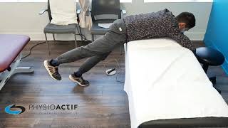
The McKenzie approach is the most studied and effective method for lumbar disc herniations with radiculopathy. This approach is based on identifying a preferential direction of movement, which is the movement that centralizes or reduces your symptoms. For the majority of posterior or posterolateral disc herniations, which account for 90% of cases, repeated lumbar extension is the preferential direction. Repeated extension movements mechanically reduce the disc herniation. They shift the nucleus pulposus forward and relieve nerve compression. You practice preferential direction exercises frequently, every 2 hours during the acute phase.
Neural mobilization aims to restore the normal gliding of nerves within their tissue interfaces. In cases of lumbar disc herniation, the nerve root can become adherent or hypersensitive. Our physiotherapists use specialized nerve treatment techniques with controlled movements. We gradually apply tension to the nervous system. These neural gliding techniques improve nerve mobility while reducing inflammation.
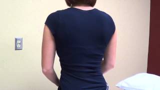
Manual therapy complements the active approach by normalizing segmental joint mobility. Our therapists apply precise joint mobilizations and manipulations to reduce protective muscle spasms. Grade I-II vertebral mobilizations produce pain-relieving effects without excessive mechanical stress on the herniated disc.
Progressive strengthening and lumbar stabilization become a priority once radicular symptoms centralize. Our program of stabilizing muscle exercises develops endurance and motor control of the deep core muscles, including the transverse abdominis and multifidus. Typical progression starts with low-load static exercises like planks and glute bridges. It advances to moderate-load dynamic exercises such as bird-dog and dead bug. It culminates in high-load functional strengthening exercises.
A Cochrane systematic review of 31 randomized controlled trials reveals that 80-90% of patients with lumbar disc herniation and radiculopathy significantly improve with conservative treatment, including physiotherapy. A prospective cohort study following 370 patients with MRI-confirmed disc herniation reports that 73% experience complete or near-complete resolution of symptoms with conservative treatment at 12 weeks.
Physiotherapy is recognized as an effective treatment for lumbar disc herniation, with results supported by scientific research.
Studies show a 75-85% success rate for treating lumbar disc herniation with physiotherapy. The combination of therapeutic exercises, manual therapy, and education proves particularly effective in reducing pain and improving function.
The effectiveness of treatment depends on several factors: how early you seek consultation (earlier = better results), consistency with home exercises, the size of the herniation, the presence of neurological symptoms, and the duration of symptoms. A comprehensive evaluation allows for tailoring the treatment to your specific situation.
Most patients notice improvement within the first 4-6 weeks of treatment, with complete resolution typically achieved in 12-16 weeks.
Are you suffering from a lumbar disc herniation? Book an appointment for a comprehensive evaluation and a personalized treatment plan.
Lumbar disc herniations can naturally resorb over a period of 3 to 12 months. This process involves inflammatory reactions and phagocytosis, which is the elimination of disc material by immune cells. Large herniations such as extrusions and sequestrations show greater resorption than small protrusions.
Serial imaging studies demonstrate that the majority of lumbar disc herniations decrease or disappear completely over time. A meta-analysis of 11 studies including 650 patients with MRI-documented disc herniations reveals that approximately 66% of herniations show a reduction in size on follow-up MRI. Among these, 43% show complete or near-complete resorption.
Biological mechanisms of resorption primarily involve inflammatory and immunological processes. When the herniated nucleus pulposus protrudes through the annulus fibrosus, it comes into contact with the vascular and immune systems for the first time. This exposure triggers an inflammatory response. Macrophages, giant cells, and neovascularization infiltrate around the lumbar disc herniation. They gradually phagocytose the herniated disc material. Paradoxically, the inflammation that initially causes pain ultimately contributes to the elimination of the herniation.
The timeline of resorption varies depending on the type and size of the lumbar disc herniation. Sequestrations, which are completely separated disc fragments, show the fastest resorption rates, reaching up to 75-100% within 6-12 months. Extrusions resorb in approximately 60-80% of cases over the same period. Protrusions show the lowest rates, approximately 40-50% within 12-24 months.
The role of physiotherapy during the natural resorption process is to manage symptoms, maintain function, and prevent detrimental compensatory adaptations during healing. We cannot directly accelerate biological resorption, but we improve the biomechanical and neurophysiological context for recovery. Patients informed about the high probability of spontaneous resorption show less anxiety. They demonstrate better treatment adherence and achieve better clinical outcomes.
We consider surgery in cases of progressive neurological deficit with worsening muscle weakness, cauda equina syndrome, which is a surgical emergency involving loss of bladder control, or failure of conservative treatment after 6-12 weeks with persistent functional limitations. Approximately 5-10% of lumbar disc herniation cases require surgery.
Cauda equina syndrome represents the only true surgical emergency. This syndrome results from a massive central lumbar disc herniation that simultaneously compresses several cauda equina nerve roots. It produces a classic triad: saddle anesthesia with loss of sensation in the perineum and around the anus, urinary retention or incontinence, and bilateral lower limb weakness. The syndrome requires surgical decompression within 24-48 hours to minimize the risk of permanent neurological sequelae. Fortunately, this syndrome occurs in less than 1-2% of lumbar disc herniations.
Need professional advice?
Our physical therapists can assess your condition and provide you with a personalized treatment plan.
Make an appointmentProgressive motor neurological deficit constitutes a semi-urgent surgical indication. If you experience muscle weakness that objectively worsens over several weeks despite conservative treatment, we must consider surgical decompression within 2-4 weeks.
Relative indications for surgery primarily include the failure of well-conducted conservative treatment. This includes the persistence of severe and disabling radicular pain after 6-12 weeks of appropriate physiotherapy treatment, as well as major functional limitations preventing work or essential daily activities. The 6-12 week timeframe is not arbitrary. The majority of patients who will improve with conservative treatment show signs of improvement within this timeframe.
Surgical outcomes are generally favorable in the short term. A meta-analysis of 64 studies including 13,055 patients reports that 78% of patients achieve good to excellent results after microdiscectomy. When comparing surgery and conservative treatment long-term over 2-4 years of follow-up, randomized controlled studies reveal similar functional and pain outcomes between the groups. Surgery primarily offers faster relief, not a superior final outcome.
Preventing a lumbar disc herniation relies on maintaining spinal flexibility, core strength with deep stabilizing muscles, appropriate lifting mechanics by keeping the load close to the body and using the legs while avoiding combined flexion-rotation, and correct posture. Regular movement breaks, ergonomic adjustments, and a healthy body weight reduce risks.
Biomechanical training and prevention techniques
Biomechanical training is the cornerstone of prevention. Safe lifting techniques include several essential principles. Keep the load close to your body to reduce leverage and lumbar stress. Primarily use your leg muscles. Avoid combined flexion-rotation movements under load. Distribute loads symmetrically. Pre-contract your abdominal muscles before lifting using the bracing technique.
General physical conditioning significantly reduces the risk of lumbar disc herniation. A balanced exercise program includes several components. Cardiovascular training maintains nutrient supply to the disc via diffusion. Core and lower limb muscle strengthening increases load-bearing capacity. Flexibility stretches maintain normal joint ranges of motion. Regular exercise improves disc hydration. Movement creates compression-decompression cycles that pump nutrients into the avascular disc.
Several lifestyle modifications contribute to prevention. Maintaining a healthy body weight is important because each kilogram of excess body weight increases disc load during daily activities. Smoking cessation offers significant benefits as smoking accelerates disc degeneration, reduces nutrient supply via vasoconstriction, increases enzymes that degrade the disc matrix, and impairs tissue repair. Adequate hydration supports overall disc health. Balanced nutrition provides the necessary nutrients for tissue maintenance.
Occupational ergonomic considerations are particularly important for workers in physically demanding jobs. Reorganize tasks to minimize repetitive lifting. Use mechanical aids such as carts and dollies. Practice task rotation to vary physical stresses. Adjust work height to avoid excessive bending. For office workers, adjustments include a chair with adjustable lumbar support, correct desk and screen height, and regular breaks from sitting with short movement breaks every 30-45 minutes.
Secondary prevention after an episode of lumbar disc herniation requires special attention. The recurrence rate can range from 5-15% within the first two years. A long-term maintenance program includes lumbar stabilization exercises 2-3 times per week and maintains continuous biomechanical awareness.
What activities should be modified with a disc herniation?
Temporarily avoid prolonged sitting, which can increase intradiscal pressure by 40-90%, heavy lifting with loads exceeding 5-10 kg initially, repeated bending, and high-impact activities during acute phases. Gradual return to activities follows symptom centralization and improved tolerance.
Activity modification follows a fundamental principle: temporarily avoid or minimize positions and movements that increase intradiscal pressure and exacerbate symptoms. Simultaneously maintain a general activity level as normal as possible. We contraindicate complete bed rest except in the most severe cases. You should limit it to a maximum of 1-2 days, as prolonged rest delays recovery.
Prolonged sitting represents the most problematic activity. Sitting, especially with lumbar flexion in a slumped posture, increases intradiscal pressure by 40-90% compared to standing. It pushes the nucleus pulposus backward. Modification strategies include limiting continuous sitting periods to 20-30 minutes followed by standing or walking breaks, using lumbar support to maintain natural lumbar lordosis, adjusting chair height to allow knees to be slightly lower than hips, and considering a sit-stand workstation.
Lifting activities require substantial modifications during the acute phase of a lumbar disc herniation. Initially, during the first 2-4 weeks, completely avoid lifting loads greater than 5-10 kg. Avoid any activity involving simultaneous flexion, rotation, and load. As your symptoms centralize, gradually reintroduce lifting. Start with light loads using perfect technique. Gradually increase the weight by 10-20% per week only if the technique remains correct and symptoms do not worsen.
Certain sports should be temporarily avoided during the acute phase. Racket sports with explosive rotations like tennis and squash, golf, contact sports like hockey and football, high-impact running, and lumbar flexion exercises such as full sit-ups and abdominal crunches should be temporarily set aside.
Other activities are generally well tolerated. Walking is an excellent basic exercise. Swimming, particularly backstroke and freestyle, is suitable, but avoid breaststroke if bending your lower back worsens your symptoms. Stationary cycling with an upright posture and resistance training for the upper and lower body, avoiding heavy axial loads, can be continued.
Your return to activities should be guided by two key principles. Symptom centralization means that your pain should not spread to other areas. Latency refers to the time between an activity and the onset of symptoms, which should gradually increase.
Get expert care for your lumbar disc herniation.Our physiotherapists at Physioactif specialize in evidence-based treatment for lumbar disc herniations.2 We can help you avoid surgery and return to your normal activities through personalized rehabilitation programs. Our programs target your specific lumbar disc herniation pattern, symptoms, and functional goals.
Our approach integrates the most effective techniques validated by scientific research. We use the McKenzie assessment to identify your preferred direction of movement. We apply neural mobilization to restore normal nerve gliding. We perform manual therapy to improve joint mobility. We develop personalized progressive exercise programs to restore strength, endurance, and motor control.
Beyond immediate symptom relief, we equip you with the knowledge and skills needed to manage your condition long-term and prevent it from returning. Our educational approach helps you to understand what causes your pain. You will understand the natural healing process of your lumbar disc herniation. You will learn effective self-management strategies and lifestyle changes that protect your spine.
Evidence strongly supports a conservative approach led by physiotherapy. Research shows that about 80-90% of patients with a lumbar disc herniation can avoid surgery with appropriate physiotherapy treatment. Even for those who do have surgery, post-operative rehabilitation improves functional results and helps reduce the chances of the problem returning.
References
- Brinjikji W, Luetmer PH, Comstock B, et al. Systematic literature review of imaging features of spinal degeneration in asymptomatic populations. AJNR Am J Neuroradiol. 2015;36(4):811-816.
- Fardon DF, Williams AL, Dohring EJ, et al. Lumbar disc nomenclature: version 2.0. Spine J. 2014;14(11):2525-2545.
- Bogduk N. Clinical and Radiological Anatomy of the Lumbar Spine. 6th ed. Edinburgh: Elsevier; 2022.
- Lurie JD, Tosteson TD, Tosteson AN, et al. Surgical versus nonoperative treatment for lumbar disc herniation: eight-year results for the spine patient outcomes research trial. Spine. 2014;39(1):3-16.
- Zhong M, Liu JT, Jiang H, et al. Incidence of spontaneous resorption of lumbar disc herniation: a meta-analysis. Pain Physician. 2017;20(1):E45-E52.
- Zhong M, Liu JT, Jiang H, et al. Incidence of spontaneous resorption of lumbar disc herniation: a meta-analysis. Pain Physician. 2017;20(1):E45-E52.
- Jordan J, Konstantinou K, O'Dowd J. Herniated lumbar disc. BMJ Clin Evid. 2011;2011:1118.
- Brinjikji W, Luetmer PH, Comstock B, et al. Systematic literature review of imaging features of spinal degeneration in asymptomatic populations. AJNR Am J Neuroradiol. 2015;36(4):811-816.
- Adams MA, Roughley PJ. What is intervertebral disc degeneration, and what causes it? Spine. 2006;31(18):2151-2161.
- McGill SM, Marshall L, Andersen J. Low back loads while walking and carrying. Ergonomics. 2013;56(2):293-302.
- Nachemson AL. Disc pressure measurements. Spine. 1981;6(1):93-97.
- Kelsey JL, Githens PB, O'Conner T, et al. Acute prolapsed lumbar intervertebral disc. Spine. 1984;9(6):608-613.
- Battié MC, Videman T, Gibbons LE, et al. Determinants of lumbar disc degeneration. Spine. 1995;20(24):2601-2612.

Other conditions
Hip osteoarthritis is the normal wear and tear of the hip joint. It is often said that osteoarthritis is the wear and tear of the cartilage between our bones. That is true, but it involves more than just the cartilage. Cartilage is a tissue that acts as a cushion between the surfaces of our bones and allows our joints to glide smoothly and move fluidly.
This is normal wear and tear of the knee joint. It’s often said that osteoarthritis is the wearing down of the cartilage between our bones. That’s true, but it’s more than just the cartilage. Cartilage is a tissue that acts as a cushion between the surfaces of our bones and allows our joints to glide smoothly and move fluidly.
It is an inflammation of the subacromial bursa in the shoulder joint.
A bursa is a small, thin sac filled with fluid that is found in many of the body's joints. This small sac acts as a cushion within the joint and lubricates the structures that are subject to increased friction.
It is a tissue that surrounds the shoulder and helps keep the shoulder bone in place within the joint. The capsule helps keep the joint stable.
Neck pain is a general term used to describe pain in the neck that has no specific cause, such as an accident or a sudden awkward movement. Neck pain is therefore synonymous with “my neck hurts, and nothing in particular happened.”
In both types of injury, pain is felt in the neck and then radiates into the arm, or vice versa.
Make an appointment now
We offer a three-pronged quality assurance approach: optimized treatment time, a second opinion from a physical therapist, and ongoing expertise to ensure effective care tailored to your needs.

Customer satisfaction is our top priority
At Physioactif, excellence guides everything we do, but our patients are the best ones to tell you about it. Take a look at their verified reviews to get a real sense of their experience.
Discover our physical therapy clinics
We have locations in several areas to better serve you.
Blainville
190 Bas-de-Sainte-Thérèse Road, Suite 110,
Blainville, Quebec
J7B 1A7
Located in Blainville, near Rosemère, the Physioactif clinic is easily accessible to residents of the area and the surrounding communities
Laval
3224 Jean-Béraud Ave., Suite 220, Laval,
QC H7T 2S4
Located in Chomedey, in the heart of Laval, the Physioactif clinic is easily accessible to people in the area
Montreal
8801 Lajeunesse Street,
Montreal,
QC H2M 1R8
Located in Ahuntsic, near Villeray, the Physioactif clinic is easily accessible to residents of both neighborhoods
Saint-Eustache
180 25th Avenue, Suite
201 Saint-Eustache
QC J7P 2V2
Located in Saint-Eustache, the Physioactif clinic is easily accessible to residents of the area and the surrounding communities
Vaudreuil
21 Cité-des-Jeunes Boulevard, Suite 240,
Vaudreuil-Dorion, Quebec
J7V 0N3
Located in Vaudreuil-Dorion, the Physioactif clinic is easily accessible to people in the area
Make an appointment now
