Lumbar Spinal Stenosis

You might notice your legs feeling heavy, numb, or painful when you walk. Interestingly, the pain lessens when you lean forward or push a grocery cart. Perhaps you've been told you have a 'narrowing of the spinal canal' in your back. This diagnosis can sound alarming, especially if you're over 50.
Good news: Spinal stenosis can be managed very effectively with physiotherapy.¹ You are not condemned to stop moving; there are effective strategies to maintain your mobility and quality of life. What science tells us about spinal stenosis:- Spinal stenosis is a normal part of spinal aging; it's not a 'serious disease'.
- Leaning forward opens the spinal canal and relieves pressure on the nerves, which is why walking while bent forward helps.
- Unlike a herniated disc, stenosis develops slowly over several years.
- Lumbar flexion and strengthening exercises are often very effective.
This guide explains how to maintain an active life despite stenosis. To understand the broader context of lower back pain, consult our complete guide to back pain.
What is Lumbar Spinal Stenosis?
Lumbar spinal stenosis is the narrowing of the spinal canal (the tunnel for the nerves) in the lower back. This narrowing compresses the nerves, causing pain, numbness, and weakness in the legs, especially in adults over 50.
The spinal canal is the space that runs through your entire spine.¹ It contains the spinal cord and nerve roots. It's like a bony tunnel that protects your nerves. When this space becomes smaller, it's called spinal stenosis or central stenosis. The narrowing can occur at different points in the lower back.¹ Stenosis is one of several conditions that affect the lumbar region. If you suffer from back pain, discover our comprehensive back pain program.
There are two types of lumbar stenosis. Congenital stenosis (present from birth) is rare; you are born with a naturally narrower canal. Acquired stenosis develops over time.² This is the most common form and is part of the normal aging process of the spine.
Unlike a herniated disc, which occurs suddenly, stenosis develops gradually over several years. A herniation typically compresses a single nerve abruptly, whereas stenosis often affects both sides. Consult our complete guide to lumbar disc herniation to understand this important difference in treatment.
What Causes the Spinal Canal to Narrow?
Stenosis develops from age-related changes. These include disc degeneration (the cushions wear out), osteoarthritis of the joints, and thickening of the ligaments. These changes gradually reduce the space available for the nerves until the compression causes symptoms.
The process leading to stenosis is part of the aging of the spine.³ It often begins with disc dehydration. Discs are the cushions between the vertebrae, like thick sponges between each bone. When discs lose their thickness, the vertebrae move closer together. The joints at the back of the spine then bear more load.³
Degenerative Processes Causing Lumbar Stenosis
| Degenerative Process | Mechanism | Effect on the canal |
|---|---|---|
| Disc degeneration | Disc Dehydration and Loss of Height | Vertebrae Moving Closer and Joint Overload |
| Facet joint arthritis | Wear and Tear of Joint Cartilage | Bone spur formation that narrows the canal |
| Ligament thickening | Hypertrophy of the Ligamentum Flavum (Yellow Ligament) | Compression at the back of the canal |
| Osteophyte Formation | Bone Spurs for Stabilization | Narrowing of the Space Available for Nerves |
This overload triggers a complex degenerative process. Lumbar osteoarthritis involves the wear and tear of cartilage and the formation of osteophytes (bone spurs).⁴ These arthritic changes are directly responsible for the narrowing of the canal. To understand how osteoarthritis progresses and contributes to stenosis, consult our complete guide to lumbar osteoarthritis.
Several factors increase the risk of developing stenosis. Age remains the primary factor: prevalence significantly increases after 60 years of age.¹,² Other factors include genetics and activities that involve repeated heavy lifting.²
What are the Classic Symptoms of Lumbar Stenosis?
Neurogenic claudication is the typical symptom. This involves pain, numbness, and weakness in the legs. These symptoms worsen when you walk or stand for long periods and improve when you sit or lean forward. While these symptoms can be frustrating in daily life, they are well understood and treatable. You may also experience lower back pain, balance problems, and symptoms on both sides.
When stenosis causes symptoms, pain can appear in the lower back, buttocks, or legs.¹ The pain increases when you walk. It also increases when standing for long periods. It increases when you lean your back backward. Conversely, the pain decreases when you lean forward. It lessens when sitting or lying down.¹ This pattern of relief with flexion represents a typical directional preference for stenosis, which is opposite to what is observed with herniated discs. Going up stairs is easier than going down them.¹
The 'shopping cart sign' is very telling. Many patients report that leaning on a shopping cart relieves their symptoms.⁵ This relief occurs because the slightly bent-forward position increases the space in the spinal canal. Many patients also find they can ride a stationary bike without problems, but walking is difficult.⁵ This is because the seated position on a bike opens up the canal.
Difference Between Neurogenic and Vascular Claudication
| Characteristic | Neurogenic Claudication (Stenosis) | Vascular Claudication (Arterial) |
|---|---|---|
| Cause | Nerve compression in the canal | Lack of oxygen in the muscles |
| Trigger | Walking and standing | Walking only |
| Relief | Sitting or leaning forward | Stopping while standing (rest) |
| Relief time | 5-10 minutes | 1-2 minutes |
| Bilateral/Unilateral | Often bilateral (both legs) | Often unilateral (one leg) |
It's important to differentiate between these two types of claudication (pain while walking).² With vascular claudication, symptoms improve when you stop walking, and you can remain standing. With neurogenic claudication, however, you need to sit down or lean forward to find relief.²
Some individuals with stenosis experience no back pain at all;¹ all their symptoms are in their legs. Numbness or tingling sensations can indicate nerve irritation.¹ While these symptoms might resemble radiculopathy, the distribution and behavior of the pain are different. Learn how to distinguish these conditions in our complete guide to lumbar radiculopathy.
It's important to know that stenosis can be completely asymptomatic.¹ MRI studies have shown that many people have a narrowing of the spinal canal without experiencing any symptoms. This distinction between imaging findings and actual symptoms is crucial. Treatment should always be based on your real symptoms, not solely on what an MRI shows.²
10 Quick Tips for Understanding Your Pain
The ones that have made the biggest difference in my patients' lives. 1 a day, 2 minutes.
How is lumbar spinal stenosis diagnosed?
Diagnosis is made by combining your clinical history of neurogenic claudication, a physical examination, and an MRI. Your physiotherapist or doctor will look for symptoms that worsen with back extension (leaning backward) and improve with flexion (leaning forward). While an MRI will reveal any narrowing of the spinal canal, your functional symptoms are more important than the severity seen on the imaging.
The diagnosis is primarily made based on your symptom history and a clinical examination.¹ Your physiotherapist or doctor will look for the typical pattern: leg symptoms triggered by walking and relieved by sitting. The physical examination includes tests where leaning backward reproduces your symptoms,⁵ and leaning forward provides relief.
Clinical tests to identify spinal stenosis
Provocation tests used in assessment:- Standing Extension Test: You lean backward while standing. This action reproduces or worsens your leg symptoms.
- Walking Test: This test assesses how far you can walk before symptoms appear. Some patients can walk 50-100 meters, while others can walk several hundred meters.
- Mobility Assessment: We examine the movement of your lower back and how your nerves glide.
- Strength Tests: We assess your muscle strength and stability to pinpoint any specific weaknesses.
A physiotherapy assessment also looks at your joint mobility and the quality of your movements.¹ These findings help us identify the issues contributing to your limitations and guide your treatment plan.
Imaging and MRI Severity Grade
In some cases, you may need imaging tests.¹ X-rays or an MRI can show the severity of the stenosis. Magnetic Resonance Imaging (MRI) directly visualizes the spinal canal and measures its diameter. Radiologists often categorize stenosis as mild, moderate, or severe.²
It's important to understand that what appears on imaging isn't always directly related to your symptoms.¹ The connection between how severe the narrowing looks and your actual symptoms is often modest.² Some patients with severe stenosis on an MRI experience very few symptoms, while others with moderate stenosis are significantly limited. If your MRI shows considerable stenosis, don't be discouraged; it's not a definitive diagnosis of your condition. This discrepancy means that treatment decisions should always be based on your actual symptoms and how they limit you.²
Imaging becomes important when surgery is being considered, as it helps identify which areas need decompression. However, for physiotherapy, a functional assessment is more useful than the exact severity shown on an MRI.²
How Does Physiotherapy Manage Lumbar Stenosis?
Physiotherapy focuses on flexion exercises, manual therapy, postural training, and cardiovascular conditioning. The goal is to maximize the space available for your nerves and improve your overall function. We emphasize active strategies that help you maintain your mobility and quality of life, even with the spinal canal narrowing.
Your physiotherapist will conduct a thorough assessment, evaluating your joint mobility, how your nerves glide, and your strength.¹ This detailed assessment helps identify specific issues and allows us to create a personalized treatment plan. Physiotherapy offers several effective approaches to manage lower back pain and improve your function.
Treatment is built upon several key principles. Firstly, flexion exercises are designed to increase space within the spinal canal.⁵ These exercises include stretches such as knee-to-chest and pelvic tilts. Stationary cycling is especially beneficial, as it provides cardiovascular conditioning while maintaining a forward-bent spine.⁵
Secondly, muscle strengthening is crucial in managing stenosis. Strengthening and muscle endurance exercises improve your movement control and help protect your spine.¹ Focusing on the deep core stabilizing muscles is especially important. These muscles reduce the strain on arthritic areas and improve your overall posture.
Thirdly, manual therapy provides significant relief. Joint mobilizations and manipulations help restore spinal movement and reduce mechanical stress.¹ Nerve treatment techniques improve how nerves glide, allowing them to move more freely within the spinal canal. This combined approach maximizes the space available for your nerves.
Your physiotherapist will teach you how to properly manage your daily activities.¹ You'll receive advice on posture and movement. Understanding your condition empowers you to make informed choices, helping you identify which activities and positions worsen or relieve your symptoms.⁵
It's important to understand that stenosis cannot be cured; it's a wear-and-tear process that doesn't reverse. However, treatment can completely eliminate your symptoms,¹ even if the stenosis still appears on imaging. This fact emphasizes the importance of focusing on your function and quality of life.
What is the Prognosis for Lumbar Stenosis?
Rest assured: lumbar stenosis typically progresses slowly. Many patients maintain good function through conservative management. While the anatomical narrowing doesn't reverse, symptoms can be effectively managed with physiotherapy. Only 10 to 15% of patients require surgery over a 5-year follow-up period.
The natural progression of stenosis is generally benign, advancing slowly over several years.² Contrary to some beliefs, stenosis doesn't always lead to worsening symptoms. Follow-up studies show that many patients remain stable, and some even improve over time, especially when they participate in a physiotherapy program.²
The success rates for conservative treatment are encouraging. Between 85% and 90% of patients can manage their condition without surgery.² By combining activity modifications, postural strategies, and physical conditioning, they can continue their important daily activities despite the narrowing of the spinal canal.
It's important to know that 90% of lower back pain episodes resolve within 6 to 12 weeks,¹ and 50% resolve within 1 to 2 weeks. While stenosis is a chronic condition, periods of worsening often follow this natural pattern of resolution,¹ especially when combined with appropriate management.
Several factors influence the prognosis. Patients who maintain regular physical activity tend to have better outcomes,² as do those who apply the strategies learned in physiotherapy. It's important to avoid the fear of movement. However, if muscle weakness worsens, it might suggest a more guarded prognosis, and in such cases, surgery could be necessary.²
The majority of patients can maintain a high quality of life.² The goal isn't to eliminate all symptoms, but rather to manage them to a level that allows you to continue your important activities and maintain your independence.
Need professional advice?
Our physical therapists can assess your condition and provide you with a personalized treatment plan.
Make an appointmentWhen Is Surgery Recommended for Spinal Stenosis?
Decompression surgery (laminectomy, where a part of the bone is removed) is considered for severe neurogenic claudication that limits walking to less than 200 meters. It is also considered for progressive neurological deficits, or if conservative treatment has failed after 3 to 6 months of structured physiotherapy. Surgery offers good results for appropriate patients.
Surgical indications are clearly defined. The presence of cauda equina syndrome (compression of nerves at the base of the spine) is a surgical emergency.² This syndrome involves loss of bladder or bowel control, numbness in the genital and anal region, and severe weakness in both legs. While it's important to be aware of these warning signs, please be reassured that this syndrome is extremely rare, occurring in less than 1% of cases.¹
Serious symptoms to watch for include:¹
- Severe weakness or paralysis in one or both legs
- Significant loss of sensation in one or both legs
- Recent Loss of Balance While Walking
- Significant and new incoordination
- New difficulties holding your urine or stool
- Loss of sensation in the genital area
For non-urgent cases, surgery is typically considered when claudication is so severe that it significantly limits your quality of life,² even after trying conservative treatment. An adequate trial generally involves 3 to 6 months of physiotherapy with activity modifications.² If, after this period, you can only walk 50 to 100 meters, a surgical consultation is appropriate, especially if your muscle weakness is also progressing.²
The most common type of surgery is decompressive laminectomy.² The surgeon removes a part of the vertebra and other structures that are compressing the spinal canal, such as bone spurs (osteophytes) and thickened ligaments. This decompression creates more space for the nerves. In some cases, spinal fusion (joining two vertebrae together) is also performed if there is instability.²
Surgical outcomes are generally good for suitable patients, with approximately 70 to 80% reporting significant improvement in their leg symptoms.² However, surgery does carry risks, including infection, bleeding, and cerebrospinal fluid leakage. In rare cases, neurological complications can occur.² Even after successful surgery, physiotherapy rehabilitation is essential to help regain strength, mobility, and optimal function.²
How to modify daily activities for stenosis?
Modifications include using shopping carts or walkers for support, taking frequent sitting breaks during walks, sleeping with elevated knees, and maintaining good ergonomics at work. These strategies help you manage symptoms while maintaining your independence and activity levels.
Several practical strategies can be implemented immediately:
Practical modifications per activity:- When grocery shopping: Use the cart for support and lean slightly forward¹
- Walking: Take short, regular sitting breaks (every 100-200 meters, depending on your tolerance)¹
- Standing still: Limit static standing time; sit down every 20 minutes¹
- Prolonged standing: Place one foot in front of the other and shift your weight slightly¹
- Cooking: Place one foot inside the cabinet in front of you to slightly flex your back¹
- Sleeping: Lie on your side with a pillow between your knees, or on your back with pillows under your knees¹,⁵
For walking, adopt a strategy of taking breaks. Instead of walking a long distance all at once, plan your route with places where you can sit down. This approach often allows you to cover a much greater total distance.
If you must stand, micro-movements can help. Shifting your weight slightly changes the pressure on your spine, which can delay the onset of symptoms.¹
Using assistive devices can be very beneficial. A walker or cane allows you to walk further.⁵ They provide support and encourage a slightly forward-leaning posture. If you hesitate to use these aids, know that they are a tool for freedom, not a sign of weakness. They help maintain independence and participation in valued activities.
Temporarily stop movements that cause too much pain, then gradually resume them later.¹ However, do not remain completely immobile for long periods. You will become stiffer and experience more pain afterward.¹ The balance between respecting pain and maintaining movement is crucial.
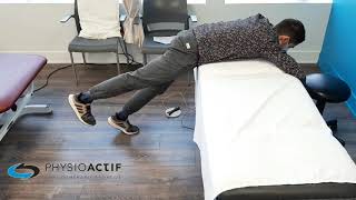
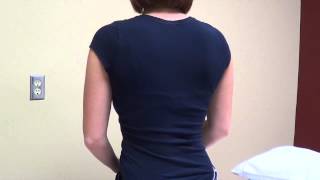
What exercises help manage stenosis symptoms?
Flexion exercises such as knee-to-chest stretches, pelvic tilts, and stationary cycling help open the spinal canal. These are combined with core strengthening and hip flexibility exercises to support spinal stability. Exercise selection is individualized based on your symptomatic response and goals.
Performing repeated back flexion movements can relieve symptoms.¹ These movements temporarily increase space in the spinal canal. For example, gently bend forward to touch your toes. Hold this position for 10 to 30 seconds.⁵ You can do this several times a day.
Stationary cycling is an excellent cardiovascular exercise option.⁵ The seated, forward-leaning position on the bike keeps the spine in flexion, allowing for aerobic conditioning. Many patients who can only walk for a few minutes can cycle for 20 to 30 minutes. A recumbent bike is often even more comfortable as it eliminates load on the spine.
Aquatic exercises also offer excellent benefits.⁵ Buoyancy in water reduces the load on the spine, allowing for movements that would be difficult on land. Water walking, aqua aerobics, and swimming are appropriate options. However, swimming on your stomach with your head out of the water should be avoided, as it forces lumbar extension.
Strengthening core muscles helps support the spine.⁵ This reduces stress on arthritic structures. Stabilization exercises, such as modified planks (on your knees if necessary), strengthen deep muscles. These exercises do not create excessive lumbar extension. They should be taught by a physiotherapist to ensure proper technique.
Hip flexibility exercises are important. Stiff hip flexors pull on the pelvis, which increases lumbar lordosis (the curve in the lower back) and can worsen stenosis.⁵ Gentle stretches for the psoas and piriformis muscles help. Hip mobilizations also help maintain mobility, allowing the spine to stay in a neutral position.
A progressive conditioning approach is essential.⁵ Begin with gentle, short-duration exercises and gradually increase their intensity and duration as tolerated. Some days will be better than others, so it's important to adjust your program based on your current symptoms. Do not push through significant pain.
If you don't see any improvement after 10 days, you should consult a physiotherapist.¹ A personalized and supervised program is generally more effective, as it can be tailored to your specific needs and individual response.
Stenosis is a classic example of a condition that responds well to flexion exercises. To better understand why and discover other exercises tailored to your pain pattern, consult our complete guide on directional preferences.
How does stenosis differ from other back conditions?
Unlike a disc herniation, which typically causes sciatica on one side, stenosis usually causes symptoms on both sides. These symptoms worsen when standing and improve when sitting. Vascular claudication, on the other hand, improves simply by stopping walking while remaining standing. These distinctions help guide the appropriate diagnosis and treatment.
Differentiating Lumbar Conditions
| Characteristic | Lumbar Stenosis | Herniated disc | Vascular Claudication | Hip osteoarthritis |
|---|---|---|---|---|
| Onset | Gradual (years) | Acute (sudden) | Gradual | Gradual |
| Typical Age | >50 years old | 30-50 years old | >60 years old | >60 years old |
| Leg Symptoms | Bilateral (both sides) | Unilateral (one side) | One or both sides | Groin and thigh |
| Worsened by | Extension, standing | Flexion, sitting | Walking | Hip internal rotation |
| Relieved by | Sitting, flexion | Extension, walking | Stopping while standing | Rest |
| Pain distribution | Vague, diffuse | Precise nerve pathway | Calf muscles | Mainly groin |
| Spinal position | Major influence | Moderate influence | No influence | No influence |
The distinction between spinal stenosis and a herniated disc is clinically important. A herniated disc usually occurs suddenly, often after a bending and twisting movement while carrying a load. It typically compresses a single nerve root on one side.⁶ This leads to classic sciatica: pain that travels down one leg along a precise path, accompanied by numbness and sometimes weakness. To better understand this radiating pain, consult our complete guide on lumbosciatica.
In contrast, stenosis develops gradually over many years. It often affects several spinal levels, causing symptoms on both sides that are typically more vague.² Stenosis pain is aggravated by extension (leaning backward) and standing. A herniated disc, however, is often worsened by flexion (leaning forward) and sitting.²
Distinguishing between neurogenic and vascular claudication is also crucial.² With vascular claudication, symptoms improve when you stop and stand still because resting muscles require less oxygen. With neurogenic claudication, you need to sit down or lean forward. It's the change in your spine's position that relieves pressure on the nerves, not simply resting your muscles. This difference in how position affects symptoms is often the simplest way to tell the two conditions apart.²
Hip osteoarthritis can also cause pain in the buttock and thigh.⁴ These pains might be mistaken for stenosis. However, hip osteoarthritis typically causes pain in the groin, which gets worse with walking and hip movements. This type of pain is not affected by the position of your spine, which helps distinguish it from neurogenic claudication.⁴
These distinctions highlight the importance of a thorough clinical evaluation. While imaging can confirm the presence of stenosis, a herniated disc, or osteoarthritis, it's your medical history and a physical examination that help determine which structure is causing your symptoms.² This, in turn, helps us decide on the most appropriate treatment.
When should you consult a physiotherapist for spinal stenosis?
You should consider physiotherapy if you experience one or more of the symptoms described, especially characteristic neurogenic claudication: leg pain that worsens with walking and improves with sitting or bending forward. You can seek physiotherapy even if your doctor hasn't yet ruled out all other potential causes.¹
You do not need a doctor's referral to see a physiotherapist.¹ In Quebec, physiotherapists are primary care professionals. They can independently assess and treat musculoskeletal conditions. If your condition requires medical attention, your physiotherapist will inform you.¹ This allows for quicker access to treatment.
However, certain warning signs require urgent medical attention. These serious symptoms include:¹
- Severe weakness or paralysis in one or both legs
- Significant loss of sensation in one or both legs
- Recent Loss of Balance While Walking
- Significant and new incoordination
- New difficulties holding your urine or stool
- Loss of sensation in the genital area
These symptoms could indicate cauda equina syndrome, which requires emergency surgery.² Fortunately, severe symptoms are present in less than 1% of low back pain cases.¹ Most of the time, even though the pain can be uncomfortable, the condition responds well to conservative management.
Early physiotherapy consultation helps establish an accurate diagnosis, rule out any warning signs, and quickly start a tailored treatment program. This can prevent your condition from worsening and improve your overall function.
Ready to effectively manage your spinal stenosis?
Our physiotherapists at Physioactif specialize in the conservative management of spinal stenosis. We develop personalized programs to maximize your function and mobility. We help you understand your condition, improve your movement strategies, and maintain your quality of life despite the narrowing of the spinal canal.
Lumbar spinal stenosis does not have to mean a life of pain and limitations. With a good understanding of your condition, tailored strategies, and an appropriate exercise program, the vast majority of patients maintain excellent function and continue to enjoy their valued activities.²
The goal is not to "cure" degenerative changes, as these are often permanent. Instead, it's about managing your symptoms and improving your functional ability despite these changes.
Don't wait until your symptoms become severe before seeking help. Early intervention often prevents the condition from worsening and helps establish lifelong management habits. Our physiotherapists will work with you to develop a personalized plan that respects your personal goals, current limitations, and your ability to commit to rehabilitation.
To schedule an appointment with one of our physiotherapists specializing in lumbar conditions, contact your nearest Physioactif clinic. Together, we can help you regain control of your spinal health and live fully despite lumbar spinal stenosis.
References
- Kreiner DS, Shaffer WO, Baisden JL, Gilbert TJ, Summers JT, Toton JF, Hwang SW, Mendel RC, Reitman CA. An evidence-based clinical guideline for the diagnosis and treatment of degenerative lumbar spinal stenosis (update). The Spine Journal. 2013 Jul 1;13(7):734-43.
- Lurie J, Tomkins-Lane C. Management of lumbar spinal stenosis. BMJ. 2016 Jan 4;352:h6234.
- Wu L, Cruz R. Lumbar spinal stenosis [Internet]. StatPearls Publishing; 2020 Sep 3 [cited 2025 Oct 11]. Available from: https://www.ncbi.nlm.nih.gov/books/NBK563269/
- Ordre professionnel de la physiothérapie du Québec. Osteoarthritis and Physiotherapy [Internet]. Montreal: OPPQ; 2024 [cited 2025 Oct 11]. Available: https://oppq.qc.ca/
- Ammendolia C, Stuber KJ, Rok E, Rampersaud R, Kennedy CA, Pennick V, Steenstra IA, de Bruin LK, Furlan AD. Nonoperative treatment for lumbar spinal stenosis with neurogenic claudication. Cochrane Database Syst Rev. 2013 Aug 30;(8):CD010712.
- Alexander CE, Varacallo M. Lumbosacral radiculopathy [Internet]. StatPearls Publishing; 2019 Mar 23 [cited 2025 Oct 11]. Available from: https://www.ncbi.nlm.nih.gov/books/NBK430837/

Other conditions
Hip osteoarthritis is the normal wear and tear of the hip joint. It is often said that osteoarthritis is the wear and tear of the cartilage between our bones. That is true, but it involves more than just the cartilage. Cartilage is a tissue that acts as a cushion between the surfaces of our bones and allows our joints to glide smoothly and move fluidly.
This is normal wear and tear of the knee joint. It’s often said that osteoarthritis is the wearing down of the cartilage between our bones. That’s true, but it’s more than just the cartilage. Cartilage is a tissue that acts as a cushion between the surfaces of our bones and allows our joints to glide smoothly and move fluidly.
It is an inflammation of the subacromial bursa in the shoulder joint.
A bursa is a small, thin sac filled with fluid that is found in many of the body's joints. This small sac acts as a cushion within the joint and lubricates the structures that are subject to increased friction.
It is a tissue that surrounds the shoulder and helps keep the shoulder bone in place within the joint. The capsule helps keep the joint stable.
Neck pain is a general term used to describe pain in the neck that has no specific cause, such as an accident or a sudden awkward movement. Neck pain is therefore synonymous with “my neck hurts, and nothing in particular happened.”
In both types of injury, pain is felt in the neck and then radiates into the arm, or vice versa.
Make an appointment now
We offer a three-pronged quality assurance approach: optimized treatment time, a second opinion from a physical therapist, and ongoing expertise to ensure effective care tailored to your needs.

Customer satisfaction is our top priority
At Physioactif, excellence guides everything we do, but our patients are the best ones to tell you about it. Take a look at their verified reviews to get a real sense of their experience.
Discover our physical therapy clinics
We have locations in several areas to better serve you.
Blainville
190 Bas-de-Sainte-Thérèse Road, Suite 110,
Blainville, Quebec
J7B 1A7
Located in Blainville, near Rosemère, the Physioactif clinic is easily accessible to residents of the area and the surrounding communities
Laval
3224 Jean-Béraud Ave., Suite 220, Laval,
QC H7T 2S4
Located in Chomedey, in the heart of Laval, the Physioactif clinic is easily accessible to people in the area
Montreal
8801 Lajeunesse Street,
Montreal,
QC H2M 1R8
Located in Ahuntsic, near Villeray, the Physioactif clinic is easily accessible to residents of both neighborhoods
Saint-Eustache
180 25th Avenue, Suite
201 Saint-Eustache
QC J7P 2V2
Located in Saint-Eustache, the Physioactif clinic is easily accessible to residents of the area and the surrounding communities
Vaudreuil
21 Cité-des-Jeunes Boulevard, Suite 240,
Vaudreuil-Dorion, Quebec
J7V 0N3
Located in Vaudreuil-Dorion, the Physioactif clinic is easily accessible to people in the area
Make an appointment now
