Low Back Directional Preference
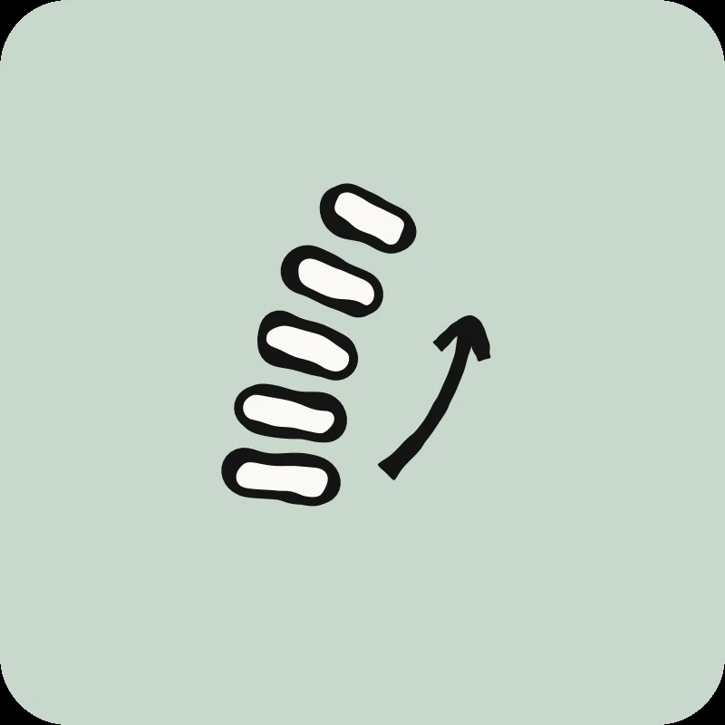

Does your back pain worsen when you're sitting? Or rather, when you're standing? The answer to this simple question could transform your recovery. Directional preference categorizes your lower back pain based on how it responds to movement and guides the selection of exercises that will truly relieve it.
Why does your pain's behavior guide treatment better than an MRI?
MRIs show abnormalities in 30% of 20-year-olds and 90% of 60-year-olds who have no pain.1 How your pain behaves in different positions guides treatment more effectively than radiological images, as these images don't always reflect the clinical reality.
Only 5 to 10% of lower back pain cases have a diagnosis identifiable through imaging.2 MRIs do not explain pain in the vast majority of cases. In contrast, movement-based classification can categorize 75% of mechanical lower back pain.3 This significant proportion demonstrates the effectiveness of the directional approach in understanding and treating your condition.
This clinical approach observes how your pain reacts when you move in different directions. It accurately identifies positions that relieve your symptoms versus those that worsen them. The McKenzie method, based on directional preference, has strong scientific evidence for reducing pain and speeding up recovery.4 Many physiotherapists use this approach to customize your exercises according to your specific pattern.
To better understand your lower back pain as a whole and discover the various possible causes, consult our complete guide to lower back pain.
What is Flexion Pain and How to Recognize It?
Flexion pain worsens with prolonged sitting, while driving, or when bending forward to pick up an object. It accounts for about 70% of mechanical lower back pain.3 An important distinguishing factor: the transition from sitting to standing occurs without major difficulty. This is precisely what differentiates it from a disc pattern, where this transition causes problems.
What are the typical symptoms of the flexion pattern?
The classic 'strained back' or 'lumbago' generally falls into this flexion pattern. It often occurs after a sudden bending movement, such as lifting a box from the floor, tying your shoes, or picking up a dropped object. The pain can be immediate or appear a few hours after the triggering activity.
People who work long hours sitting in front of a computer frequently exhibit this pattern. Maintaining a seated position for several hours increases intradiscal pressure and creates progressive tension in the posterior structures of your lumbar spine.
What exercises relieve the flexion pattern?
Seated Pelvic Tilt: Sit on the edge of your chair with your feet flat on the floor. Gently tilt your pelvis forward and backward to find the neutral position of your back, where you feel the least tension. Consciously maintain this position during your daily seated activities, whether at work, in the car, or at home.
Standing Hip Hinge: When you need to bend forward, bend at your hips rather than at your lower back. Keep your back straight and lean forward by hinging at your hips. This technique protects your back when you need to bend down to pick something up or work in a bent-over position.
Gentle Lumbar Extension: While standing, place your hands on your lower back and gently lean backward. This extension movement helps counteract the effects of prolonged flexion. Repeat this exercise several times a day, especially if you sit for long periods.
To Avoid: The exercise of bringing your knees to your chest. Although this stretch is popular, it increases the flexion load on your spine and can worsen your symptoms if you have a flexion pattern.
What is Extension Pain and How to Recognize It?
Extension pain worsens when you stand still for long periods, walk slowly (like in a shopping mall), or work with your arms overhead (painting a ceiling, putting things on high shelves). It quickly improves by sitting down or leaning on a counter to reduce lumbar lordosis. Irritated facet joints and spinal stenosis are often involved in this pattern.
What are the typical symptoms of the extension pattern?
This pattern is recognized by several distinctive characteristics:
- Pain that gradually appears while cooking standing up, especially when preparing meals that require you to remain still
- Noticeable discomfort while grocery shopping, particularly if you walk slowly while looking at shelves
- Immediate relief when you lean on a kitchen counter or a grocery cart
- Marked worsening when you work with your arms overhead, such as painting a ceiling or putting objects on high shelves
- Difficulty standing to socialize at events, with a need to sit down regularly
People with lumbar osteoarthritis often exhibit this extension pattern. Prolonged standing compresses the posterior structures of the spine, including the facet joints and the nerves exiting between the vertebrae. This compression causes the characteristic pain of the extension pattern.
People over 60 are more likely to develop this pattern due to natural degenerative changes that affect the spinal joints over time.
What exercises help relieve the extension pattern?
Standing Pelvic Tilt: While standing, gently pull your belly button towards your spine by lightly tightening your abdominal muscles. This position reduces the excessive arch in your lower back and relieves pressure on the small joints at the back of your spine, which are a source of pain. Maintain this corrected position during your prolonged standing activities.
Psoas Stretch: When the psoas muscle is tight, it increases the arch in your lower back. To stretch it, get into a lunge position with one knee on the floor. Move your pelvis forward while keeping your torso upright. You should feel a stretch at the front of the hip of your back leg.
Deep Abdominal Strengthening: Lying on your back with your knees bent, pull your belly button towards your spine by contracting your deep abdominal muscles. Hold this contraction for 10 seconds while breathing normally. Strong abdominal muscles help control an excessive arch in your lower back.
Glute Strengthening: Lying on your back with your knees bent and feet flat on the floor, squeeze your glutes and slightly lift your pelvis off the floor. Strong glutes stabilize the pelvis and reduce the tendency for excessive lower back extension during standing activities.
Absolutely Avoid: Repeated lower back extensions (such as the "Superman" exercise or cobra pose) and all exercises in a strongly arched position that increase compression on the structures at the back of your spine.
10 Quick Tips for Understanding Your Pain
The ones that have made the biggest difference in my patients' lives. 1 a day, 2 minutes.
What is Mixed Pain or Disc-Related Pain?
Mixed or disc pain is recognized by a major distinguishing characteristic: a marked difficulty transitioning from sitting to standing. You may need to walk hunched over for a few steps, sometimes even holding onto furniture, before you can fully straighten up. This is the most important differentiating characteristic of this pattern compared to others.
How do you distinguish disc pain from other patterns?
The key distinguishing characteristic is a difficult sit-to-stand transition. If you have difficulty straightening up after sitting for a while, and you need to walk a few steps hunched over before you can stand upright, you likely have a disc or mixed pain pattern.
Other important specific characteristics:
- Both bending forward (flexion) and arching backward (extension) worsen your symptoms, unlike pure patterns.
- Pain varies considerably throughout the day (often worse in the morning upon waking).
- Initial acute phase: arching your lower back is very limited and painful.
- Subacute phase (after a few days): walking gradually starts to relieve symptoms.
- Sudden movements or prolonged sustained positions worsen the condition.
This pattern often suggests a disc issue where the inner core of the intervertebral disc puts pressure on surrounding structures. A herniated disc is a classic example of this pattern, although not all cases of mixed pain pattern are necessarily herniated discs.
What is sacroiliac pain?
Sacroiliac pain differs from the three classic directional patterns in several distinctive ways:
Need professional advice?
Our physical therapists can assess your condition and provide you with a personalized treatment plan.
Make an appointment- Pain localized on one side, often precisely at the visible dimple in the lower back.
- Rapid onset of pain regardless of the position adopted (sitting, standing, lying down).
- Walking does NOT relieve the pain, unlike disc pain in the subacute phase.
- Pain often triggered by asymmetrical movements or prolonged positions on one leg.
- Marked local tenderness when the sacroiliac joint is touched (palpated).
This condition requires a specific evaluation and different treatment techniques than the classic lumbar directional patterns.
How to Identify Your Pattern in 5 Simple Questions?
Three key questions quickly help identify your pain pattern: Is your pain worse when sitting or standing? Does it get better when you change positions? Do you have trouble straightening up after sitting for a while?
Detailed self-assessment questions:- Is your pain worse after sitting for a long time or standing for a long time?
- Does your pain significantly improve when you change positions?
- Do you have difficulty straightening up completely after sitting?
- Does walking relieve or worsen your pain?
- Is your pain localized to one specific side, or is it more central and spread out?
This classification helps guide your initial self-treatment quickly. However, a complete professional evaluation is still recommended to confirm your pattern and customize your treatment plan for the best results.
When should you see a professional?
Consult a physiotherapist if your pain lasts more than 6 to 8 weeks without significant improvement, if this is your second episode of low back pain, or if the pain travels down your leg past the knee. Fortunately, red flags requiring urgent medical attention are present in less than 1% of cases.7
Statistics show that about 90% of acute low back pain resolves naturally within 6 to 8 weeks, with or without specific treatment. However, the recurrence rate reaches 40 to 70% in the following year.8 Identifying your pattern from the first episode and learning the right exercises significantly reduces the risk of future relapse.
Early consultation also helps prevent the development of inappropriate muscle compensations and avoidance behaviors that can unnecessarily prolong your pain episode.
What are the red flags that require immediate consultation?
Consult a doctor immediately or go to the emergency room if you experience any of the following symptoms:
- Loss of bladder control (new urinary incontinence) or bowel control (fecal incontinence)
- Progressive numbness in the genital or perineal region (saddle anesthesia)
- Muscle weakness that rapidly progresses in both legs simultaneously
- High fever accompanied by severe and unexplained low back pain
- Significant unintentional weight loss associated with back pain
- History of cancer with new low back pain
- Pain that constantly worsens despite rest and appropriate treatments
These rare symptoms can indicate serious conditions such as cauda equina syndrome, a spinal infection, or a pathological fracture, which require urgent medical intervention.
If your low back pain travels down your leg and causes numbness or tingling, consult our detailed article on lumbosciatica to better understand this specific condition.
How does Physioactif evaluate and treat your directional pattern?
Our specialized physiotherapists systematically evaluate your directional pattern from the very first assessment session. They confirm your pattern with standardized repeated movement tests and immediately customize your exercises based on your individual response to the different movements tested.
The complete assessment includes:- Detailed questions about how your pain behaves in various positions and activities
- Repeated movement tests in different directions to clearly identify your directional preference
- Careful observation of your sit-to-stand transition and dynamic posture
- Identifying exercises and positions that provide immediate relief during the session
- Assessment of your range of motion and current functional limitations
- Neurodynamic tests if pain radiates into the leg
Research shows that patients with a clearly identifiable directional preference achieve better clinical outcomes.4 Early and accurate identification of your pattern leads to significantly faster and more lasting recovery. Self-management strategies taught from the start also significantly reduce the risk of future recurrence.
Our physiotherapists also use specific manual techniques tailored to your pattern: joint mobilizations, myofascial release, and neuromuscular techniques that effectively complement your personalized exercise program.
To learn more about the complete physiotherapy assessment process and the different treatment approaches available, consult our detailed guide on physiotherapy for back pain.
References
- Brinjikji W, et al. Systematic literature review of imaging features of spinal degeneration in asymptomatic populations. AJNR Am J Neuroradiol. 2015;36(4):811-816.
- Deyo RA, Weinstein JN. Low back pain. N Engl J Med. 2001;344(5):363-370.
- Fritz JM, et al. Subgrouping patients with low back pain. Phys Ther. 2007;87(4):441-454.
- May S, Donelson R. Evidence-informed management of chronic low back pain with the McKenzie method. Spine. 2008;33(22):E887-E900.
- Delitto A, et al. Low back pain clinical practice guidelines. Phys Ther. 2012;92(4):e1-e51.
- Long A, et al. Does it matter which exercise? Spine. 2004;29(23):2593-2602.
- Henschke N, et al. Prevalence of and screening for serious spinal pathology. BMJ. 2009;339:b3829.
- da Silva T, et al. Risk of recurrence of low back pain. Spine. 2017;42(13):1003-1009.
Videos in this category
Other conditions
Hip osteoarthritis is the normal wear and tear of the hip joint. It is often said that osteoarthritis is the wear and tear of the cartilage between our bones. That is true, but it involves more than just the cartilage. Cartilage is a tissue that acts as a cushion between the surfaces of our bones and allows our joints to glide smoothly and move fluidly.
This is normal wear and tear of the knee joint. It’s often said that osteoarthritis is the wearing down of the cartilage between our bones. That’s true, but it’s more than just the cartilage. Cartilage is a tissue that acts as a cushion between the surfaces of our bones and allows our joints to glide smoothly and move fluidly.
It is an inflammation of the subacromial bursa in the shoulder joint.
A bursa is a small, thin sac filled with fluid that is found in many of the body's joints. This small sac acts as a cushion within the joint and lubricates the structures that are subject to increased friction.
It is a tissue that surrounds the shoulder and helps keep the shoulder bone in place within the joint. The capsule helps keep the joint stable.
Neck pain is a general term used to describe pain in the neck that has no specific cause, such as an accident or a sudden awkward movement. Neck pain is therefore synonymous with “my neck hurts, and nothing in particular happened.”
In both types of injury, pain is felt in the neck and then radiates into the arm, or vice versa.
Make an appointment now
We offer a three-pronged quality assurance approach: optimized treatment time, a second opinion from a physical therapist, and ongoing expertise to ensure effective care tailored to your needs.

Customer satisfaction is our top priority
At Physioactif, excellence guides everything we do, but our patients are the best ones to tell you about it. Take a look at their verified reviews to get a real sense of their experience.
Discover our physical therapy clinics
We have locations in several areas to better serve you.
Blainville
190 Bas-de-Sainte-Thérèse Road, Suite 110,
Blainville, Quebec
J7B 1A7
Located in Blainville, near Rosemère, the Physioactif clinic is easily accessible to residents of the area and the surrounding communities
Laval
3224 Jean-Béraud Ave., Suite 220, Laval,
QC H7T 2S4
Located in Chomedey, in the heart of Laval, the Physioactif clinic is easily accessible to people in the area
Montreal
8801 Lajeunesse Street,
Montreal,
QC H2M 1R8
Located in Ahuntsic, near Villeray, the Physioactif clinic is easily accessible to residents of both neighborhoods
Saint-Eustache
180 25th Avenue, Suite
201 Saint-Eustache
QC J7P 2V2
Located in Saint-Eustache, the Physioactif clinic is easily accessible to residents of the area and the surrounding communities
Vaudreuil
21 Cité-des-Jeunes Boulevard, Suite 240,
Vaudreuil-Dorion, Quebec
J7V 0N3
Located in Vaudreuil-Dorion, the Physioactif clinic is easily accessible to people in the area
Make an appointment now
