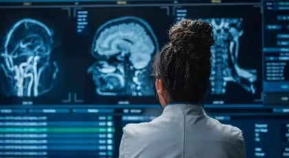

You are in pain and would like to have an X-ray or an MRI (magnetic resonance imaging), a test that produces detailed images of the inside of the body. You hope to understand what is causing the pain. This is a natural desire. Imaging can provide a helpful answer, but its role depends on the suspected problem and treatment decisions.
An imaging test is not always necessary to begin treatment. For low back pain, the healthcare professional’s evaluation first looks for warning signs: a major accident, fever, loss of strength or sensation, or unexplained weight loss. In the absence of these signs, routine immediate imaging generally does not improve pain or a person’s ability to perform daily activities.1
In a study that followed 3,369 people for six years, most lumbar changes seen on MRI were not associated with future pain intensity. Some associations were found, but they were generally weak.2 The imaging findings should therefore be interpreted in conjunction with your symptoms and the physical examination.
10 Quick Tips to Understand Your Pain
One tip a day for 10 days to help you better understand your pain. About a 2-minute read.
- Changes seen on an MRI of the back are common in people without pain.3
- A change in the image may occur without causing any pain.3
- The way an MRI result is explained can affect a patient’s level of concern.4
- For low back pain without any concerning signs, routine immediate imaging generally does not improve treatment outcomes.1
This guide helps you understand when imaging is truly useful. To learn more about the relationship between what is seen and what is felt, read our article on osteoarthritis and pain.
Do imaging tests always show where it hurts?
Imaging does not always reveal the cause of pain: some changes also occur in people who are not in pain. However, an abnormality may contribute to the symptoms. Its significance depends on the overall evaluation.
When we have an injury or pain, it's in our nature to want to know the exact structure that is hurting us.
Not knowing what the problem is can be worrisome, such as when you're trying to figure out the cause of a water leak in the basement.
X-rays, ultrasounds, CT scans, and MRIs show different parts of the body. An ultrasound uses sound waves to produce images; a CT scan uses X-rays. These tests can reveal a fracture or other injury, but an image does not directly measure the pain a person feels.
For back, knee, or shoulder pain that is not the result of an accident or impact, guidelines generally advise against routine imaging at the start of treatment. The healthcare professional can often determine the appropriate initial treatment based on the interview and physical examination.5 For low back pain without a specific cause, routine imaging offers no significant benefit and may lead to unnecessary interventions.6
Does an imaging test change the treatment plan?
An imaging test may alter the treatment plan if its results reveal a problem that requires a different course of treatment. However, its routine use for lower back pain and certain knee problems generally provides little or no benefit in terms of pain, daily activities, satisfaction, or quality of life.7 The plan is based primarily on the physical therapy evaluation and on the specific question that the imaging is intended to answer.
The decision depends on what the review can actually change. These two situations illustrate the difference:
| Situation | The Role of Imaging |
|---|---|
| Lower back pain with no cause for concern | The evaluation may make it possible to begin treatment without immediately obtaining an image. |
| Suspected fracture or other specific problem | An appropriate image can guide the medical diagnosis and influence treatment. |
A study examined the timing of imaging in 782 people who had already been referred to an orthopedist or neurosurgeon—doctors who, among other things, evaluate conditions that may require surgery. Overall treatment outcomes were similar whether imaging—such as an MRI or CT scan—was performed early or reserved for needs that arose later. However, the early-imaging group showed a slight improvement in certain outcomes at eight and twenty-four months.8 The choice therefore also depends on the clinical situation.
An unnecessary imaging test can lead to additional tests or treatments with no expected benefit.6 How the results are explained also matters. In a trial involving 44 people with back pain, one group received a factual explanation of the MRI report. The other group received a reassuring explanation presenting the results as normal changes. After six weeks, the first group had a more negative perception of their back, less improvement in pain, and more difficulty with daily activities.4 This small trial suggests that results should be explained in context. It does not show that any imaging worsens back pain.
Interpretations of MRI results can also vary. In one study, a 63-year-old woman with back pain and symptoms in one leg underwent MRIs at ten different centers over a three-week period. None of the 49 distinct findings appeared in all ten reports.9 This case illustrates variability among reports; it does not measure the reliability of all exams for all patients.
Three limitations help explain why a picture isn't always enough.
What are the three concepts that explain this discrepancy?
Three limitations explain this discrepancy: the image does not directly measure pain, the body changes with age, and some abnormalities exist without symptoms.
First, pain is a personal experience. A medical image does not directly show what you are feeling. The healthcare professional listens to your description and correlates it with the physical examination.10, 11 Just as with sadness, one can observe symptoms without actually experiencing what the other person is going through. A normal image therefore does not invalidate your pain.
Second, the body changes with age. The discs—the cushions between the bones of the spine—and the joints can change in appearance. These changes become more common with age, even in the absence of pain.3 Osteoarthritis refers to changes affecting the entire joint; its presence on an image does not necessarily explain the symptoms.
In a study involving 3,110 people with no history of back pain, researchers estimated the prevalence of changes by age. At age 40, the estimated prevalence of disc degeneration was 68%. This term refers, among other things, to changes in the disc’s water content and height. At age 30, the estimated prevalence of disc bulging was 40%: in this condition, the disc protrudes more significantly beyond its normal space.3 These estimates do not mean that all changes observed in a person experiencing pain are insignificant.
Third, an abnormality can exist without causing pain. Some people have osteoarthritis in their lower back without experiencing pain. This does not rule out the possibility that osteoarthritis contributes to symptoms in another person.3 For back pain, physical therapy and exercise are among the treatments that can help people resume activities more comfortably. The British National Health Service (NHS) describes these treatment options for back pain.
For knee osteoarthritis, a 2024 review found short-term improvements in pain and physical function with exercise. Compared to no treatment, usual care, or limited advice, the average improvement was about 13 points out of 100 for pain and 13 for physical function. The uncertainty surrounding the averages did not allow for the conclusion that the benefits consistently exceeded the thresholds chosen for a significant change.12 These results pertain to the knee; they do not directly measure the effect of exercise on lumbar osteoarthritis.
In another study, 98 adults without back pain underwent an MRI. The researchers found bulging in 52% of the participants, protrusion in 27%, and extrusion in 1%. Protrusion and extrusion are two forms of a herniated disc, in which the disc’s contents locally extend beyond their normal space.13 These findings do not automatically indicate the cause of pain in another person.
The figures above refer to the lower back.3 MRI changes are also present in the necks of people without symptoms. A review of cervical MRI studies confirms their presence, while also finding that certain changes are more common in people with a disease of the spinal cord—the bundle of nerves that connects the brain to the body.14 Evaluation therefore remains necessary.
When is medical imaging truly useful?
Medical imaging is useful when it is used to identify a specific cause or provides information that may change the course of treatment. A suspected fracture, a serious accident, a possible infection, or a decision to undergo surgery may warrant it. Progressive weakness, spreading numbness, fever, or unexplained weight loss require evaluation.6 The healthcare professional determines the next steps based on these signs before selecting a treatment plan.
The physical therapist looks for factors that may warrant an imaging test in order to determine the best treatment plan. In Quebec, a physical therapist who holds the required certification may order an X-ray following an injury to muscles, bones, or joints that occurred within the past 72 hours. The physical therapist must be the first professional to assess the injury—without prior medical consultation—and must comply with the ordering criteria. For other imaging tests, the physical therapist will refer you to a physician. The interpretation of X-rays is reserved for physicians. Rules of the Quebec Professional Order of Physical Therapy.
For non-traumatic pain in the lower back, knee, and shoulder, guidelines generally recommend imaging only for suspected specific or serious problems. Imaging may also be considered if initial non-surgical treatment is ineffective and if the results are likely to change the course of treatment.5 Age-related changes must be interpreted in conjunction with the symptoms.3
Don’t delay seeing a doctor while waiting for test results. Back pain, a sensation of heat or cold, fever, chills, or general malaise are all reasons to seek urgent medical attention. Severe pain that begins suddenly, or pain that worsens rapidly, also warrants urgent care. Unexplained weight loss also requires medical attention.
Go to the emergency room if your back pain is accompanied by any of the following symptoms:
- Pain, tingling, weakness, or numbness in both legs.
- Loss of sensation around the genitals or anus.
- New difficulty urinating or leakage of urine or stool.
- Changes in genital sensation during sexual intercourse, difficulty achieving or maintaining an erection, or an inability to reach orgasm.
- Chest pain.
- Back pain began after a serious accident.
Warning signs described by the NHS.
Is a herniated disc detected on an imaging test a death sentence?
A herniated disc detected on imaging alone does not determine the severity of your pain or your functional abilities. In a study of 56 people with a lumbar herniated disc, the relative size of the herniation was not associated with leg pain or difficulties with daily activities. People with urgent nerve involvement affecting bladder or bowel function were excluded.15
Hernias can also change over time as seen on imaging. In a Danish study, 140 herniated discs in 106 people were monitored between the ages of 41 and 49. Over eight years, 65% of the hernias remained stable, 17.5% decreased in size, 12.5% increased in size, and 5% fluctuated. These changes in size alone do not account for the progression of symptoms.16 To learn more about this condition and possible treatments, visit our page on lumbar disc herniation.
To learn more
Physical therapist Alexis Gougeon hosts the podcast *Parle-moi de santé*. In Episode 27, he discusses medical imaging with physical therapist Tatiana Vukobrat and family physician René Wittmer.
Episode #27 of Parle-moi de santé (medical imaging)
Listen to Episode 27 on the podcast's website.
Need professional advice?
Our physical therapists can assess your condition and recommend a personalized treatment plan.
Make an appointmentSources:
Links open in a new tab.
- Chou R, Fu R, Carrino JA, Deyo RA. Imaging strategies for low-back pain: systematic review and meta-analysis. Lancet. 2009;373(9662):463-72. (Back to sections: 1, 2)
- Kasch R, Truthmann J, Hancock MJ, Maher CG, Otto M, Nell C, et al. Association of Lumbar MRI Findings with Current and Future Back Pain in a Population-Based Cohort Study. Spine (Phila Pa 1976). 2022;47(3):201-211. (Back to section: 1)
- Brinjikji W, Luetmer PH, Comstock B, Bresnahan BW, Chen LE, Deyo RA, et al. Systematic literature review of imaging features of spinal degeneration in asymptomatic populations. AJNR Am J Neuroradiol. 2015;36(4):811-6. (Back to sections: 1, 2, 3, 4, 5, 6, 7)
- Rajasekaran S, Dilip Chand Raja S, Pushpa BT, Ananda KB, Ajoy Prasad S, Rishi MK. The effects of an MRI report on catastrophizing in patients and surgeons and the benefits of “clinical reporting”: results from an RCT and blinded trials. Eur Spine J. 2021;30(7):2069-2081. (Back to sections: 1, 2)
- Cuff A, Parton S, Tyer R, Dikomitis L, Foster N, Littlewood C. Guidelines for the use of diagnostic imaging in musculoskeletal pain conditions affecting the lower back, knee, and shoulder: A scoping review. Musculoskeletal Care. 2020;18(4):546-554. (Back to sections: 1, 2)
- Chou R, Qaseem A, Owens DK, Shekelle P, Clinical Guidelines Committee of the American College of Physicians. Diagnostic imaging for low back pain: advice for high-value health care from the American College of Physicians. Ann Intern Med. 2011;154(3):181-9. (Back to sections: 1, 2, 3)
- Karel YH, Verkerk K, Endenburg S, Metselaar S, Verhagen AP. Effect of routine diagnostic imaging for patients with musculoskeletal disorders: A meta-analysis. Eur J Intern Med. 2015;26(8):585-95. (Back to section: 1)
- Gilbert FJ, Grant AM, Gillan MG, Vale LD, Campbell MK, Scott NW, et al. Low back pain: influence of early MRI or CT on treatment and outcome—a multicenter randomized trial. Radiology. 2004;231(2):343-51. (Back to section: 1)
- Herzog R, Elgort DR, Flanders AE, Moley PJ. Variability in diagnostic error rates at 10 MRI centers performing lumbar spine MRI examinations on the same patient within a 3-week period. Spine J. 2017;17(4):554-561. (Back to section: 1)
- Wideman TH, Edwards RR, Walton DM, Martel MO, Hudon A, Seminowicz DA. The Multimodal Assessment Model of Pain: A Novel Framework for Further Integrating the Subjective Pain Experience into Research and Practice. Clin J Pain. 2019;35(3):212-221. (Back to section: 1)
- Raja SN, Carr DB, Cohen M, Finnerup NB, Flor H, Gibson S, et al. The revised International Association for the Study of Pain definition of pain: concepts, challenges, and compromises. Pain. 2020;161(9):1976-1982. (Back to section: 1)
- Lawford BJ, Hall M, Hinman RS, Van der Esch M, Harmer AR, Spiers L, et al. Exercise for osteoarthritis of the knee. Cochrane Database Syst Rev. 2024;12(12):CD004376. (Back to section: 1)
- Jensen MC, Brant-Zawadzki MN, Obuchowski N, Modic MT, Malkasian D, Ross JS. Magnetic resonance imaging of the lumbar spine in people without back pain. N Engl J Med. 1994;331(2):69-73. (Back to section: 1)
- Banerjee A, Mowforth OD, Nouri A, Budu A, Newcombe V, Kotter MRN, et al. The Prevalence of Degenerative Cervical Myelopathy-Related Pathologies on Magnetic Resonance Imaging in Healthy/Asymptomatic Individuals: A Meta-Analysis of Published Studies and Comparison to a Symptomatic Cohort. J Clin Neurosci. 2022;99:53-61. (Back to section: 1)
- Dunsmuir RA, Nisar S, Cruickshank JA, Loughenbury PR. No correlation was found between the proportional size of a prolapsed intervertebral disc and disability or leg pain. Bone Joint J. 2022;104-B(6):715-720. (Back to section: 1)
- Kjaer P, Tunset A, Boyle E, Jensen TS. Progression of lumbar disc herniations over an eight-year period in a group of adult Danes from the general population—a longitudinal MRI study using quantitative measures. BMC Musculoskeletal Disorders. 2016;17:26. (Back to section: 1)
- Jarvik JG, Deyo RA. Diagnostic evaluation of low back pain with an emphasis on imaging. Ann Intern Med. 2002;137(7):586-97.
- Murphy L, Schwartz TA, Helmick CG, Renner JB, Tudor G, Koch G, et al. Lifetime risk of symptomatic knee osteoarthritis. Arthritis Rheum. 2008;59(9):1207-13.
- van Tulder MW, Assendelft WJ, Koes BW, Bouter LM. Spinal radiographic findings and nonspecific low back pain. A systematic review of observational studies. Spine (Phila Pa 1976). 1997;22(4):427-34.
Customer satisfaction is our top priority
At Physioactif, excellence guides everything we do, but our patients are the best ones to tell you about it. Take a look at their verified reviews to get a real sense of their experience.









Discover our physical therapy clinics
We have locations in several areas to better serve you.
Blainville
190 Bas-de-Sainte-Thérèse Road, Suite 110,
Blainville, Quebec
J7B 1A7
Located in Blainville, near Rosemère, the Physioactif clinic is easily accessible to residents of the area and the surrounding communities
Boucherville
690 Rue de Montbrun, Suite S,
Boucherville, Quebec
J4B 8H2
Located in Boucherville, the Physioactif clinic is easily accessible to people in the area
Laval
3224 Jean-Béraud Ave., Suite 220, Laval,
QC H7T 2S4
Located in Chomedey, in the heart of Laval, the Physioactif clinic is easily accessible to people in the area
Montreal
8801 Lajeunesse Street,
Montreal,
QC H2M 1R8
Located in Ahuntsic, near Villeray, the Physioactif clinic is easily accessible to residents of both neighborhoods
Saint-Eustache
180 25th Avenue, Suite
201 Saint-Eustache
QC J7P 2V2
Located in Saint-Eustache, the Physioactif clinic is easily accessible to residents of the area and the surrounding communities
Vaudreuil
21 Cité-des-Jeunes Boulevard, Suite 240,
Vaudreuil-Dorion, Quebec
J7V 0N3
Located in Vaudreuil-Dorion, the Physioactif clinic is easily accessible to people in the area
Make an appointment now