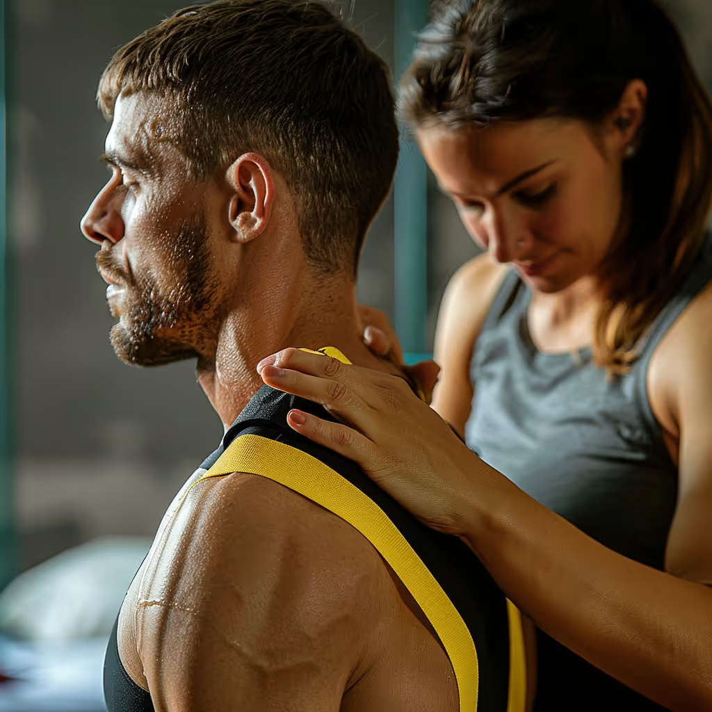


The cervical spine is the upper part of your spine. Understanding this anatomy helps put neck pain into context, without attributing the symptoms to a single structure.
Your neck performs a remarkable feat every day. The junction between the skull and the first cervical vertebrae helps stabilize the head while allowing you to turn, tilt, and bend it. The ligaments in this region combine a high degree of mobility with significant stability and protection for the nervous and vascular structures.1
What makes the cervical spine unique compared to the rest of the spine?
The cervical spine differs from other regions of the spine in that it is exceptionally mobile and has smaller vertebrae.
The cervical vertebrae are generally smaller than the thoracic and lumbar vertebrae. Their shape and characteristics vary depending on the cervical level.
10 Quick Tips to Understand Your Pain
One tip a day for 10 days to help you better understand your pain. About a 2-minute read.
Typical cervical vertebrae (C3 to C6) share unique characteristics:
| Characteristic | Description | Function |
|---|---|---|
| Small vertebral body | Smaller than the chest and lower back vertebrae | Supports the head and transfers loads to the rest of the spine |
| Triangular opening | Triangular opening for the spinal cord | The space through which the spinal cord passes |
| Split bony projection | Tip divided into two | Muscle attachment points |
| Transverse openings | Holes in the side projections | Typical course of the vertebral arteries from C6 to C1 |
To understand how the cervical region fits into the overall spine, consult our guide on spinal anatomy.
How are the cervical vertebrae arranged?
The seven cervical vertebrae extend from C1, below the skull, to C7, above the thorax. C1 and C2 form the upper cervical region and have distinct shapes. C3 through C6 are often described as the typical cervical vertebrae. C7, on the other hand, exhibits characteristics that mark the transition to the thoracic region.
What makes C1 (the atlas) so special?
The atlas (C1) is the first cervical vertebra. It doesn't have a vertebral body or a spinous process like other vertebrae. It forms a bony ring that directly supports the skull. Its name comes from the titan Atlas in Greek mythology, who carried the world on his shoulders.
The atlas has a unique structure:
- Ring shape: Unlike other vertebrae, C1 is essentially a ring of bone
- Lateral masses: A total of two bony structures, one on each side, that articulate with the skull and C2
- No vertebral body: C1 lacks a vertebral body, unlike typical cervical vertebrae
- Anterior and posterior arches: Connect the lateral masses into a complete ring
The atlanto-occipital joint (C0-C1) connects the base of the skull to the first cervical vertebra and primarily allows the head to tilt forward and backward.
What is the role of C2 (the axis)?
The axis (C2) is a cervical vertebra that has a bony projection called the dens, or odontoid process.
The dens is a tooth-shaped projection that rises upward behind the anterior arch of C1, within the ring formed by the atlas.
The atlantoaxial joint connects C1 to C2 and allows for most of the head's rotation. The dens forms the pivot around which the atlas rotates.
What distinguishes C7 from the other cervical vertebrae?
C7, also known as the vertebra prominens, has a longer, non-bifid spinous process that you can easily feel at the base of your neck. This vertebra marks the transition between the cervical and thoracic regions of the spine.
When you tilt your head forward and touch the base of your neck, the bony bump you feel is likely C7. It is a useful anatomical landmark for healthcare professionals.
How do cervical intervertebral discs work?
The cervical discs distribute the load among the vertebrae while allowing the neck to move and helping to maintain its stability. They begin between C2 and C3; there is no disc between C1 and C2. They are generally thicker at the front than at the back.
The height of the intervertebral discs and vertebral bodies can be assessed on imaging studies. This measurement alone is not sufficient to predict a person’s mobility or symptoms.
Each cervical disc consists primarily of two parts:2
- Nucleus pulposus: A water-rich, gel-like center that contributes to the disc's load-bearing capacity.
- Fibrous ring (annulus fibrosus): The outer layers that surround the nucleus and are rich in collagen, a protein that forms strong fibers
The unciform processes are small bony projections on the lateral edges of the cervical vertebral bodies.
If you want to learn more about cervical disc herniations, consult our complete guide on cervical disc herniation.
What are the important joints of the cervical spine?
The atlanto-occipital, atlanto-axial, facet, and uncovertebral joints connect the bones of the skull and neck to one another. The facet joints are located at the back of the vertebrae, while the uncovertebral joints—also known as Luschka's joints—are located on the sides of the cervical vertebral bodies.
How do facet joints guide neck movements?
The surfaces of the facet joints glide against one another at the back of the vertebrae. Their shape, along with the discs and capsules surrounding them, guides movement between the vertebrae and limits the range of motion. The orientation of the facets varies depending on the level of the neck, but their slope alone does not determine the movements.3, 4
These joints are covered with cartilage and surrounded by a joint capsule, a fibrous sheath that encloses the joint.Cervical osteoarthritis can affect the facet joints in particular. For this specific component, the review found no positive association between signs of osteoarthritis and neck pain.5
What are the unvertebral joints (Luschka's joints)?
The uncovertebral joints are unique to the cervical region. They are located between the uncinate processes, which extend from the lateral edges of the vertebral bodies of C3 through C7, and the adjacent vertebrae.
The anatomy of the intervertebral spaces varies from person to person and depending on the cervical level.
How do the spinal cord and nerves pass through the cervical region?
The spinal cord runs through the canal formed by the vertebrae, while the cervical nerves exit the spinal column through openings on its sides. These cervical nerves emerge at different levels of the cervical region.
How are the cervical nerves numbered?
There are eight pairs of cervical nerves, numbered C1 through C8. Nerves C1 through C7 emerge above the vertebra of the same number. C1 passes between the skull and the atlas. The C8 nerve emerges between C7 and T1, the first thoracic vertebra. Therefore, there is a C8 nerve, but no C8 vertebra.6
Which areas do the cervical roots C5 through C8 innervate?
A cervical nerve root refers here to the initial portion of a cervical spinal nerve as it exits the spinal cord. The symptoms associated with a cervical nerve root sometimes follow a rough distribution pattern. These areas overlap and are not sufficient to identify the affected level. In a study of C6 or C7 compressions confirmed by magnetic resonance imaging (MRI), the areas of decreased sensation overlapped almost completely, which calls for caution during examination.7 The C5 through C8 lines in the table below are approximate and overlapping landmarks; no single zone, muscle, or reflex is sufficient on its own to localize a nerve root.8
The table describes the areas where a nerve root can affect sensation, muscle strength, or a reflex. To test a reflex, the healthcare professional taps a tendon lightly with a hammer and observes the involuntary contraction of the muscle. The biceps reflex is tested on the front of the elbow, the triceps reflex on the back, and the brachioradialis reflex on the forearm near the wrist. These tests complement other observations.9
| Nerve root | Approximate Sensitive Marker | Approximate muscle landmark | Approximate reference point |
|---|---|---|---|
| C5 | Outer surface of the shoulder and arm | Deltoid and biceps: Raise your arm and bend your elbow | Biceps |
| C6 | Thumb side of the forearm, thumb, and index finger | Biceps and muscles that extend the wrist | Biceps and brachioradialis |
| C7 | The area in the middle of the forearm toward the thumb, index finger, and middle finger | Triceps and wrist-flexor muscles | Triceps |
| C8 | Forearm on the little-finger side, ring finger, and little finger | Small muscles of the hand and muscles that flex the fingers | No typical tendon reflex specific to C8 |
In cervicobrachialgia or cervical radiculopathy, pain and other symptoms may radiate into the arm, but their distribution varies and is not sufficient to pinpoint a specific nerve root.10 A narrowing of the spinal canal, as in cervical stenosis, can also affect these nerve structures.
Which muscles control neck movements?
The deep flexors, the sternocleidomastoid muscles, the scalene muscles, and the posterior muscles work together to move the neck. The anterior muscles are located at the front of the neck; the posterior muscles are at the back. The flexors help bend the neck or head forward. The extensors help straighten them or tilt them backward.
What are the anterior muscles of the neck?
The anterior neck muscles include the deep flexors, located near the front of the cervical spine, and superficial muscles, such as the sternocleidomastoid.
Deep Cervical Flexors:- The longus colli, or long neck muscle, is a deep muscle located at the front of the cervical spine.
- The longus capitis, or long muscle of the head, is a deep muscle that connects the upper part of the cervical spine to the base of the skull.
- The rectus capitis anterior, or anterior small straight muscle of the head, connects the first cervical vertebra to the base of the skull.
According to the assessment, physical therapy for neck pain may include exercises tailored to the patient’s symptoms, abilities, and goals.
Sternocleidomastoid (SCM):It is a visible neck muscle that forms a slanted band on each side. It attaches to the sternum and the clavicle at the bottom, and to the mastoid process at the top—the bony protrusion of the skull located behind the ear. The SCM helps rotate and tilt the head. The two SCM muscles also work together to bend the head forward.11 The SCM may be contracted during torticollis, though this contraction is not necessarily the cause of the condition.12
Scalene Muscles:The anterior, middle, and posterior scalene muscles connect the cervical vertebrae to the first two ribs. They are involved in neck movements and help lift the ribs during deep inhalation.
What are the posterior muscles of the neck?
The posterior neck muscles are arranged in layers, from those closest to the skin to those closest to the vertebrae.
Trapezius (Upper Part):This large, diamond-shaped muscle extends from the base of the skull to the middle of the back. Its upper portion attaches primarily to the occipital bone, the nuchal ligament, and the clavicle; it does not attach directly to the cervical vertebrae. Working in front of a screen or living with neck pain does not, in and of itself, mean that the symptoms can be attributed to the upper trapezius.
We often hear about “text neck”—the idea that looking at your phone can damage your neck. However, when researchers photographed the neck posture of a large group of teenagers, they found no link to pain or headaches.13 This is a cross-sectional study, which observes a group at a single point in time and therefore cannot establish cause and effect. It focuses solely on 17-year-olds and, on its own, does not allow us to draw conclusions about all adults.
10 Quick Tips to Understand Your Pain
One tip a day for 10 days to help you better understand your pain. About a 2-minute read.
The suboccipital muscles include:
- Posterior Great Straight Muscle of the Head (rectus capitis posterior major)
- Small posterior straight muscle of the head (rectus capitis posterior minor)
- Superior Oblique Muscle of the Head (obliquus capitis superior)
- Inferior oblique muscle of the head (obliquus capitis inferior)
The suboccipital muscles are located at the base of the skull. Their presence in this region is not sufficient to explain cervicogenic headaches.
Deep Extensor Muscles (Cervical Multifidi):These small muscles connect several cervical vertebrae and help straighten the neck.
Which ligaments stabilize the cervical spine?
The longitudinal, yellow, and nuchal ligaments, as well as the ligaments of the C1-C2 region, help stabilize the cervical spine. Some are present in several regions of the spine; others are found primarily in the upper cervical region.
Which ligaments run the entire length of the spine?
The anterior and posterior longitudinal ligaments run along most of the length of the spine. The yellow ligaments connect the laminae of adjacent vertebrae—that is, the posterior bony portions of their vertebral arches; in the neck, the nuchal ligament is also present.
Anterior Longitudinal Ligament:The anterior longitudinal ligament runs along the front of the vertebral bodies to the sacrum. It limits excessive backward movement.
Posterior Longitudinal Ligament:The posterior longitudinal ligament runs along the back of the vertebral bodies, inside the canal that contains the spinal cord. It helps support the spine when it bends forward.
Ligamentum Flavum (Yellow Ligament):The yellow ligaments connect the laminae of adjacent vertebrae. Their elastic fibers allow them to stretch and then return to their original shape during movement.14
Nuchal Ligament:It is an extension of the supraspinous ligament, specific to the neck. It extends from the external occipital protuberance to C7 and attaches to the cervical spinous processes.
Which ligaments are specific to C1-C2?
The junction between the skull, C1, and C2 includes several ligaments that differ from those at other cervical levels.
Transverse Ligament of the Atlas:The transverse ligament of the atlas holds the odontoid process of C2 against the anterior arch of C1. It passes behind this bony projection and attaches to the sides of the atlas.
Tectorial Membrane:The tectorial membrane extends the posterior longitudinal ligament toward the inner surface of the base of the skull. It attaches to the clivus, a bony surface located in front of the large opening in the skull. It helps stabilize the junction between the skull and the upper neck.15
Alar Ligaments:These two short ligaments connect the dens to the occipital condyles, the two rounded bony surfaces located at the base of the skull.
How do blood vessels pass through the neck?
The vertebral arteries usually ascend through the lateral foramina of the vertebrae from C6 to C1 and then enter the skull. Their anatomical course may vary.16
The typical course of each vertebral artery includes the following stages:
- The artery originates from a subclavian artery near the base of the neck.
- The artery usually enters a lateral opening in C6, called the transverse foramen.
- The artery runs upward through the vertebral foramina to C1.
- The artery runs around the back of the atlas and enters the skull through its large lower opening, the foramen magnum.
- The two vertebral arteries join inside the skull to form the basilar artery.17
The starting point and entry level into the vertebrae may vary. The right and left sides are not always identical. An anatomical variation does not automatically indicate a problem; imaging, when indicated, allows this pathway to be examined in its context.16
What is cervical lordosis and why is it important?
Cervical lordosis refers to the natural forward curve of the neck; the curve itself does not explain the pain.
An X-ray of the cervical curve must be interpreted in conjunction with the symptoms and physical examination; the shape of the curve alone is not sufficient to explain the pain.
In this cross-sectional study of 107 people over the age of 45, there was no significant difference in cervical curvature between the group with neck pain and the group without neck pain.18 This result does not allow us to predict a person’s prognosis based on their curvature.
The healthcare professional therefore combines the physical examination and symptoms with the imaging findings to guide treatment.
How can the cervical spine heal and adapt?
Neck muscles can become stronger with exercise, while recovery from injury varies depending on the affected structure and the clinical situation. In a trial involving women with persistent neck pain, strengthening exercises improved neck strength after one year.19 This result demonstrates the muscles’ ability to adapt; it does not guarantee the same outcome after every injury.
A study followed a group of people after a collision. Persistent neck pain was more strongly associated with psychological distress, pre-accident health, whiplash symptoms, and initial activity limitations than with most characteristics of the collision.20
The progression of symptoms and abilities varies depending on the diagnosis and the individual. Physical therapy can provide a plan tailored to your symptoms, abilities, and goals. Our page onwhiplash explains what you might experience after an accident.
If you are experiencing persistent neck pain or symptoms such as numbness or weakness in your arms, an evaluation by a physical therapist can help identify possible causes, check for signs that require medical referral, and tailor treatment options to your symptoms and goals. Visit our page on neck pain to schedule an appointment.
Need professional advice?
Our physical therapists can assess your condition and provide you with a personalized treatment plan.
Make an appointmentReferences
Links open in a new tab.
- Fiester P, Rao D, Soule E, Orallo P, Rahmathulla G. Anatomical, functional, and radiographic review of the ligaments of the craniocervical junction. Journal of Craniovertebral Junction and Spine. 2021;12(1):4-9. (Back to section: 1)
- Margetis K, Dowling TJ. Cervical Degenerative Disc Disease. StatPearls. 2025. (Back to section: 1)
- Jaumard NV, Welch WC, Winkelstein BA. Biomechanics and mechanotransduction of spinal facet joints in normal, injured, and degenerative conditions. J Biomech Eng. 2011;133(7):071010. (Back to section: 1)
- Nowitzke A, Westaway M, Bogduk N. Cervical zygapophyseal joints: geometric parameters and relationship to cervical kinematics. Clin Biomech (Bristol). 1994;9(6):342-8. (Back to section: 1)
- Bogduk N, MacVicar J. Osteoarthritis of the zygapophysial joints as a cause of back pain and neck pain: a scoping review. Pain Med. 2024;25(9):541-552. (Back to section: 1)
- 13.4 The Peripheral Nervous System - Anatomy and Physiology, 2nd ed. | OpenStax. (Back to section: 1)
- Rainville J, Laxer E, Keel J, Pena E, Kim D, Milam RA, et al. Exploration of sensory impairments associated with C6 and C7 radiculopathies. Spine J. 2016;16(1):49-54. (Back to section: 1)
- Hong CG, Nam WD. Reliability and Diagnostic Accuracy of Standard Dermatomes and Myotomes for Determining the Pathologic Level in Surgically Verified Patients With Cervical Radiculopathy. Neurospine. 2022;19(4):1006-1012. (Back to section: 1)
- Walker, HK. Deep Tendon Reflexes. In: Clinical Methods: The History, Physical, and Laboratory Examinations. 3rd ed. Boston: Butterworths; 1990. (Back to section: 1)
- Marco B, Evans D, Symonds N, Peolsson A, Coppieters MW, Jull G, et al. Determining the level of cervical radiculopathy: Agreement between visual inspection of pain drawings and magnetic resonance imaging. Pain Practice. 2022;23(1):32-40. (Back to section: 1)
- 11.3 Axial Muscles of the Head, Neck, and Back - Anatomy and Physiology, 2nd ed. | OpenStax. (Back to section: 1)
- Velickovic M, Benabou R, Brin MF. Cervical Dystonia. Drugs. 2001;61(13):1921-1943. (Back to section: 1)
- Richards KV, Beales DJ, Smith AJ, O'Sullivan PB, Straker LM. Neck Posture Clusters and Their Association With Biopsychosocial Factors and Neck Pain in Australian Adolescents. Phys Ther. 2016;96(10):1576-1587. (Back to section: 1)
- 7.3 The Vertebral Column - Anatomy and Physiology, 2nd ed. | OpenStax. (Back to section: 1)
- Lee SH, Cho TH, Kwon HJ, Hong JE, Lee YH, Yang HM. An anatomical and radiological study of the tectorial membrane and its clinical implications. Sci Rep. 2022;12(1):21480. (Back to section: 1)
- Tudose RC, Rusu MC, Hostiuc S. The Vertebral Artery: A Systematic Review and a Meta-Analysis of the Current Literature. Diagnostics. 2023;13(12):2036. (Back to sections: 1, 2)
- Sikka A, Jain A. Bilateral variation in the origin and course of the vertebral artery. Anat Res Int. 2012;2012:580765. (Back to section: 1)
- Grob D, Frauenfelder H, Mannion AF. The association between cervical spine curvature and neck pain. Eur Spine J. 2007;16(5):669-78. (Back to section: 1)
- Ylinen J, Takala EP, Nykänen M, Häkkinen A, Mälkiä E, Pohjolainen T, et al. Active neck muscle training in the treatment of chronic neck pain in women: a randomized controlled trial. JAMA. 2003;289(19):2509-16. (Back to section: 1)
- Atherton K, Wiles NJ, Lecky FE, Hawes SJ, Silman AJ, Macfarlane GJ, et al. Predictors of persistent neck pain after whiplash injury. Emerg Med J. 2006;23(3):195-201. (Back to section: 1)
Customer satisfaction is our top priority
At Physioactif, excellence guides everything we do, but our patients are the best ones to tell you about it. Take a look at their verified reviews to get a real sense of their experience.










Discover our physical therapy clinics
We have locations in several areas to better serve you.
Blainville
190 Bas-de-Sainte-Thérèse Road, Suite 110,
Blainville, Quebec
J7B 1A7
Located in Blainville, near Rosemère, the Physioactif clinic is easily accessible to residents of the area and the surrounding communities
Boucherville
690 Rue de Montbrun, Suite S,
Boucherville, Quebec
J4B 8H2
Located in Boucherville, the Physioactif clinic is easily accessible to people in the area
Laval
3224 Jean-Béraud Ave., Suite 220, Laval,
QC H7T 2S4
Located in Chomedey, in the heart of Laval, the Physioactif clinic is easily accessible to people in the area
Montreal
8801 Lajeunesse Street,
Montreal,
QC H2M 1R8
Located in Ahuntsic, near Villeray, the Physioactif clinic is easily accessible to residents of both neighborhoods
Saint-Eustache
180 25th Avenue, Suite
201 Saint-Eustache
QC J7P 2V2
Located in Saint-Eustache, the Physioactif clinic is easily accessible to residents of the area and the surrounding communities
Vaudreuil
21 Cité-des-Jeunes Boulevard, Suite 240,
Vaudreuil-Dorion, Quebec
J7V 0N3
Located in Vaudreuil-Dorion, the Physioactif clinic is easily accessible to people in the area
Make an appointment now