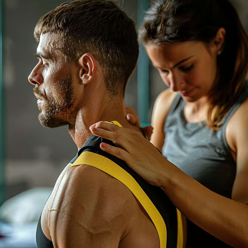


Tendons connect muscles to bones and transmit their force to produce movement. Tendon pain does not necessarily mean an irreversible injury. Exercises with progressive resistance can improve pain and function in many tendinopathies—painful tendon conditions that limit activities.1 Progression depends on the affected tendon and your tolerance. This guide explains their structure, possible causes of pain, and ways to resume your activities.
What is a Tendon and What is its Role?
A tendon is a band of strong tissue that connects a muscle to a bone and transmits muscle force to move a joint. Tendons also help maintain the position of the joints.
Tendons act like transmission cables in your body. When a muscle contracts, it pulls on the tendon. The tendon then pulls on the bone. This system allows your muscles to create precise movements.
Let's take your shoulder as an example. The tendons of the rotator cuff connect four muscles to the arm bone. When these muscles contract, their tendons pull on the bone. These movements cause your arm to rotate and lift. These tendons also help keep the arm bone in place during movement.
10 Quick Tips to Understand Your Pain
One tip a day for 10 days to help you better understand your pain. About a 2-minute read.
Some tendons store energy when they stretch and then release it when they return to their original length. This mechanism, similar to that of a spring, helps conserve muscular effort during walking and running. Its importance varies depending on the tendon and the movement.2
How are Tendons Structured?
Tendons contain collagen fibers—a strong protein—arranged in small groups that are primarily oriented in the direction of the forces pulling on the tendon. These groups of fibers transmit the forces. Cells and small blood vessels help maintain the tissue.1
Collagen fibers are made up of finer structures called fibrils. Their arrangement resembles strands of thread woven into a rope. This structure helps the tendon withstand the forces pulling on it, but excessive force can still cause it to tear.3
Tenocytes are the primary cells of the tendon. They produce and maintain the materials surrounding them, including collagen. They detect changes in load and contribute to the tissue’s response to activity.1, 3
Tendons generally receive less blood than muscles. Their blood supply varies depending on the tendon and the area being examined. Certain regions of the Achilles tendon are less vascularized—that is, they have fewer blood vessels.4 Blood supplies oxygen and nutrients. Blood flow plays a role in healing, but the number of blood vessels alone is not enough to predict the healing time.5
Why Do Tendons Become Painful?
Tendon pain can occur when changes in activity place significant strain on the tendon, but the load alone does not explain all cases. Repetitive stress, variations in load, and tissue responses may contribute to the condition.6 Pain also depends on the nervous system, which includes the nerves, the spinal cord, and the brain. The spinal cord is a cord of nervous tissue within the spine; it transmits messages between the brain and the rest of the body. Stress, fears, and life circumstances can also influence pain.7
Concerns, fears of re-injury, and how one copes with an injury can influence symptoms and the resumption of activities. This does not mean the pain is imaginary. Therefore, a change in pain does not directly reflect a change in the condition of the tendon.7
An increase in activity level that is too rapid may exceed what your body can currently handle. For example, resuming swimming three times a week after several months of inactivity is a significant change. Depending on the intensity of your workouts and your individual situation, it may be helpful to start with shorter sessions or to space them out more.
After a long period of inactivity, an intense return to exercise can be difficult to handle. The muscles and tendons have not been used in the same way for some time. A gradual return to exercise allows you to adjust your effort based on your current symptoms and abilities.8, 9
Changes in the tendon detected by ultrasound—an examination using sound waves—may occur without causing pain. However, they are not always harmless: a review of studies focusing primarily on athletes links certain abnormalities in the Achilles tendon or the patellar tendon (beneath the kneecap) to a higher risk of future pain.10 Imaging findings and symptoms must be interpreted together. The Cook and Purdam model describes different tendon responses to load but does not represent a mandatory sequence of stages for every individual.11
Seek medical attention immediately if the pain is severe or if you think you may have torn a tendon. Sudden, intense pain during an activity or following an accident may indicate a tear; you may hear a snapping sound, but this is not always the case.12
What's the Difference Between Tendinopathy and Tendinitis?
Tendinopathy refers to persistent pain in a tendon that limits its function, while tendinitis refers to inflammation of the tendon. Inflammation is a tissue reaction to irritation or injury. Tendinopathy may involve this reaction as well as changes in the tissue’s structure. The term therefore does not imply a single underlying mechanism.1
The suffix “-itis” means inflammation. The term “tendinitis” has long been used to describe many types of tendon pain. At that time, treatments often focused on relieving symptoms through rest, ice, or anti-inflammatory medications. These medications reduce certain inflammatory reactions; they do not necessarily replace a return to activity and strengthening exercises.
Microscopic examination of tendons revealed a disorganization of collagen fibers in chronic conditions. Khan’s historical review describes these changes as tendinosis.13 More recent research also describes inflammatory mediators—substances involved in tissue reactions. Their presence and collagen disorganization may coexist.1
The term “degenerative,” which is sometimes used in imaging reports, describes changes in tissue. On its own, it does not predict your pain, your ability to move, or your recovery. Your evaluation also takes your symptoms and activities into account.14, 9
Treatment depends on the symptoms and the type of injury, not just on the terms “tendinitis” or “tendinopathy.” For many types of tendinopathy, gradually increasing activity helps patients resume their normal activities. Temporary rest may be helpful in the early stages of an injury, while a tear requires a specific evaluation and specific instructions.9, 12
How do tendons heal?
Tendons repair a tear by producing repair tissue that forms a scar, and then gradually reorganizing it. This repair process involves inflammatory reactions, the production of new materials, and remodeling—that is, the reorganization of the tissue; these phases overlap.3 Recovery from persistent tendinopathy does not necessarily follow the same course. Improvement in symptoms and structural healing may occur at different rates.14
Recovery from tendinopathy can take 6 to 12 months, sometimes longer. This timeframe is not a set timeline for everyone, nor does it indicate that the tissue has fully healed.1 Your return to physical activity depends largely on the affected tendon, your symptoms, and the demands of your activities.
Progressive strengthening programs can reduce pain and improve function in several types of tendinopathy. Not all of the mechanisms underlying these benefits are fully understood. The goal of the exercises is to help you better tolerate physical activity; a completely normal imaging result is not necessary to see improvement.15, 14
Improvement after 12 weeks does not always mean a full recovery.1 In a study following 54 people with persistent mid-Achilles tendinopathy, the structure measured by ultrasound improved during an exercise program combined with an injection of platelet-rich plasma—a blood-derived preparation—or saline. At 24 weeks, statistical analysis no longer showed a clear difference between the mean values of the treated group and those of people without symptoms. However, these changes were not associated with changes in pain or functional ability.14
What is load management?
Load management adjusts the amount, intensity, and frequency of activities based on your symptoms and abilities. It aims to reduce activities that you have difficulty tolerating while maintaining those you can manage and gradually increasing your activity level.9
Physical activity includes exercise, as well as physical exertion at work, commuting, and leisure activities. A day that’s already demanding may require adjusting your planned workout.
You can temporarily reduce the duration of an activity, its intensity, or the number of repetitions. The program can then gradually increase the intensity based on your response.
Rest can alleviate symptoms, but it alone does not prepare the muscles and tendons for the demands of returning to activity. When appropriate, a substitute activity allows you to stay active with less pain.9
For example, you could shorten a painful stride or space out your running sessions. The physical therapist can also tailor strength exercises to the affected tendon. Progress depends on how you respond to the sessions, not on a set number of weeks.9
Mild pain during certain exercises may be acceptable in a diagnosed tendinopathy. Also monitor your response after the session and the following day. If pain or stiffness increases persistently, reduce the intensity and discuss making adjustments. This response helps you gauge your activity level; it does not directly measure damage to the tendon.9
Treatment trials for the Achilles and patellar tendons use pain monitoring models to guide exercises. The instructions should be tailored to your situation and explained clearly.1 Severe pain or suspicion of a tear requires prompt medical attention rather than continuing the program.12
Why isn't rest alone enough?
Rest alone does not prepare the tendon and muscles for the activities you wish to resume. A temporary reduction in activity may provide relief, but persistent tendinopathies often also require exercises and a gradual increase in load.9
First, a prolonged lack of load deprives the tendon of some of the stress that stimulates its adaptation. Inactivity reduces collagen turnover, while exercise stimulates both its production and breakdown. Both of these processes contribute to tissue maintenance.8
Second, a long period without exercise can reduce muscle strength and endurance—the muscles’ ability to sustain exertion. This is part of deconditioning, the loss of physical abilities associated with reduced activity. Returning to exercise must therefore also prepare the muscles for their usual level of exertion.9
Third, rest does not address all the factors associated with the problem. A significant increase in exertion or muscle weakness may require an adjustment to activities or strength training. Stress and fears related to movement may also warrant attention.7, 9
These recommendations apply primarily to persistent tendinopathies. A recent injury or tear may require stricter protection and rest. Follow the instructions provided after your evaluation rather than a one-size-fits-all rule that suggests rest is always harmful.12
Cortisone injections, an anti-inflammatory medication, may provide short-term relief depending on the affected tendon. For rotator cuff injuries, a review of 11 trials reports a slight reduction in pain at 4 to 8 weeks, with no clear advantage at 3 months compared to control injections consisting of saline or an anesthetic that temporarily numbs the area.16 Another review also reports short-term improvements in pain and function.17 This relief does not allow us to conclude that an injection will prevent surgery.1 In a trial of 39 men with patellar tendinopathy, cortisone and two exercise programs improved symptoms after 12 weeks. At six months, the benefits persisted in the exercise groups, while the results in the cortisone group had deteriorated.18, 1
How does progressive loading help?
Progressive loading can reduce pain and improve function in several types of tendinopathy. Progressive strengthening exercises help the muscles and tendons adapt to the demands of resuming activity. The program should take into account the affected tendon and the targeted activities.15, 9
10 Quick Tips to Understand Your Pain
One tip a day for 10 days to help you better understand your pain. About a 2-minute read.
Exercise programs can combine the following four types of exercises. Their order and difficulty are tailored to the individual; not all of them are mandatory steps.1
- Isometric exercises: You contract the muscle against resistance while keeping the joint still. In a small study of six volleyball players with patellar tendinopathy, a single session reduced pain more immediately than contractions combined with movement; the effect persisted for 45 minutes.19 However, a subsequent review combining three trials found no statistically significant difference between these two types of exercises in terms of immediate pain relief. Isometric exercises remain an option, with variable results.20, 1
- Exercises with light resistance: You move the joint against appropriate resistance. You start by performing the movement within your comfort zone, then gradually increase the difficulty.
- Slow movements with higher resistance: You increase the weight or resistance while controlling the upward and downward movements. These exercises can improve symptoms and function, particularly in certain cases of Achilles tendinopathy or patellar tendinopathy.15, 18
- High-intensity movements: If your activities require it, the program gradually reintroduces running, jumping, or changes in direction. This preparation makes the exercises more similar to the demands of the sport.9
Exercise therapy may continue beyond 12 weeks if symptoms or activity limitations persist.1 Gradual improvement may continue for several months. If the program does not produce the expected changes, a reevaluation helps adjust the workload, verify the diagnosis, and consider other treatment options.9
There is no single “right” exercise. For Achilles tendinopathy, a trial involving 58 people who had experienced pain in the middle of the tendon for more than three months compared two 12-week programs. Heavy, slow resistance training and eccentric exercises improved pain and function, with benefits lasting one year and no clear difference between the programs.15, 1 An eccentric exercise works the muscle as it lengthens, such as when you slowly lower your heel after rising onto your toes.
Targeted programs can also reduce certain problems among athletes. Among 660 elite handball players, the average proportion of participants reporting a shoulder problem during the season was 17% in the group that participated in the preventive program, compared with 23% in the control group.21 Among 652 men playing soccer at a semiprofessional level, a program to strengthen the adductors—the muscles that bring the thighs together—reduced the average proportion of groin injuries (at the crease between the thigh and the pelvis) from 21.3% to 13.5%.22 These results pertain to reported problems in these areas, not just diagnosed tendinopathies.1 For specific tendon injuries, see the guides on the shoulder and the gluteal muscles.
Do tendons truly wear out as we get older?
Tendons change with age, but these changes are not limited to wear and tear caused by movement. Their structure, cells, and ability to withstand forces can also change. Resistance exercises can still elicit adaptations in older adults.23, 24
So using a tendon doesn't automatically mean damaging it. The stresses of physical activity can trigger certain adaptations. Conversely, excessive strain or an injury can exceed the tissue's capacity to withstand it.3
The tendon remains living tissue, but not all of its components are replaced quickly. A study using carbon-14—a marker used to estimate the age of collagen—shows very little turnover in the core of a healthy Achilles tendon after growth. In affected tendons, the model estimated increased turnover over several years, sometimes before the first symptoms appeared.25, 1 Collagen turnover and improvements in strength or pain are therefore not the same phenomenon.
Research on training shows that certain properties of the tendon can change even when its central collagen undergoes little renewal. This adaptation depends, in particular, on the load and the region of the tendon being studied.23
As we age, certain cells in the tendon multiply less easily.23 A small study also compared blood flow around the Achilles tendon in 18 people, divided into three age groups. At rest, blood flow was lower in the oldest group. During exercise, the groups had similar blood flow rates.26 This finding pertains to blood flow around the Achilles tendon; it does not establish a timeline for progressing through exercises.
In older adults, resistance training can increase tendon stiffness and its resistance to deformation. This means that the tendon can stretch less under a given force, although stiffness in this context is not synonymous with pain or stiffness as experienced by the individual. These results indicate a possible adaptation that occurs with age, though they do not guarantee the same effect for everyone.23
Inactivity also affects the tendon: it reduces the renewal of its collagen. Exercise stimulates this renewal and can change the way the tendon resists forces.8 These observations support the benefits of appropriate physical activity, but they do not allow us to determine the intensity of pain based solely on tissue activity.
To maintain your fitness level, do the activities and exercises you can handle, and then gradually increase the intensity. If a tendon becomes painful, adjust the workload and have any persistent symptoms evaluated. Prolonged inactivity and a sudden return to activity are not the only options.9
To apply these principles to shoulder problems, check out our comprehensive guide to rotator cuff tendinopathy. If you have calcific tendinitis, our in-depth article on this condition explains why calcium deposits don’t mean your tendon is permanently damaged. To understand how physical therapy can help you apply these principles of progressive loading, check out our guide to physical therapy for shoulder pain.
Recovery often requires adjustments, but tendinopathy doesn’t necessarily prevent you from resuming your activities. For Achilles tendinopathy, most people recover, although some continue to experience long-term symptoms.9 A tailored program and regular reassessments allow you to track your progress and adjust your treatment as needed.
Need professional advice?
Our physical therapists can assess your condition and recommend a personalized treatment plan.
Make an appointmentWhat sources does this page draw on?
This page is based on anatomical research, treatment trials, scientific reviews, and clinical recommendations. The review by Millar and colleagues provides an overview. Items 1 and 2 refer to this same review via two different links.
Links open in a new tab.
- Millar NL, Silbernagel KG, Thorborg K, Kirwan PD, Galatz LM, Abrams GD, et al. Tendinopathy. Nat Rev Dis Primers. 2021;7(1):1. (Back to sections: 1, 2, 3, 4, 5, 6, 7, 8, 9, 10, 11, 12, 13, 14, 15, 16)
- Alexander RM. Energy-Saving Mechanisms in Walking and Running. Journal of Experimental Biology. 1991;160(1):55-69. (Back to section: 1)
- Galloway MT, Lalley AL, Shearn JT. The role of mechanical loading in tendon development, maintenance, injury, and repair. J Bone Joint Surg Am. 2013;95(17):1620-8. (Back to sections: 1, 2, 3, 4)
- Scioli MW. Achilles tendinitis. Orthop Clin North Am. 1994;25(1):177-82. (Back to section: 1)
- Tempfer H, Traweger A. Tendon Vasculature in Health and Disease. Front Physiol. 2015;6:330. (Back to section: 1)
- Magnusson SP, Langberg H, Kjaer M. The pathogenesis of tendinopathy: balancing the response to loading. Nat Rev Rheumatol. 2010;6(5):262-8. (Back to section: 1)
- Edgar N, Clifford C, O'Neill S, Pedret C, Kirwan P, Millar NL. A biopsychosocial approach to tendinopathy. BMJ Open Sport & Exercise Medicine. 2022;8(3):e001326. (Back to sections: 1, 2, 3)
- KJÆR, M. "Role of the Extracellular Matrix in the Adaptation of Tendons and Skeletal Muscle to Mechanical Loading." *Physiological Reviews*. 2004;84(2):649-698. (Back to sections: 1, 2, 3)
- de Vos RJ, van der Vlist AC, Zwerver J, Meuffels DE, Smithuis F, van Ingen R, et al. Dutch multidisciplinary guideline on Achilles tendinopathy. Br J Sports Med. 2021;55(20):1125-1134. (Back to sections: 1, 2, 3, 4, 5, 6, 7, 8, 9, 10, 11, 12, 13, 14, 15)
- McAuliffe S, McCreesh K, Culloty F, Purtill H, O'Sullivan K. Can ultrasound imaging predict the development of Achilles and patellar tendinopathy? A systematic review and meta-analysis. British Journal of Sports Medicine. 2016;50(24):1516-1523. (Back to section: 1)
- Cook JL, Purdam CR. Is tendon pathology a continuum? A pathology model to explain the clinical presentation of load-induced tendinopathy. Br J Sports Med. 2009;43(6):409-16. (Back to section: 1)
- nhs.uk. Tendonitis. 2017. (Back to sections: 1, 2, 3, 4)
- Khan KM, Cook JL, Bonar F, Harcourt P, Astrom M. Histopathology of common tendinopathies. Update and implications for clinical management. Sports Med. 1999;27(6):393-408. (Back to section: 1)
- de Jonge S, Tol JL, Weir A, Waarsing JH, Verhaar JA, de Vos RJ. The Tendon Structure Returns to Asymptomatic Values in Nonoperatively Treated Achilles Tendinopathy but Is Not Associated With Symptoms: A Prospective Study. Am J Sports Med. 2015;43(12):2950-8. (Back to sections: 1, 2, 3, 4)
- Beyer R, Kongsgaard M, Hougs Kjær B, Øhlenschlæger T, Kjær M, Magnusson SP. Heavy Slow Resistance Versus Eccentric Training as Treatment for Achilles Tendinopathy: A Randomized Controlled Trial. Am J Sports Med. 2015;43(7):1704-11. (Back to sections: 1, 2, 3, 4)
- Mohamadi A, Chan JJ, Claessen FM, Ring D, Chen NC. Corticosteroid Injections Provide Small and Transient Pain Relief in Rotator Cuff Tendinosis: A Meta-analysis. Clin Orthop Relat Res. 2017;475(1):232-243. (Back to section: 1)
- Lin MT, Chiang CF, Wu CH, Huang YT, Tu YK, Wang TG. Comparative Effectiveness of Injection Therapies in Rotator Cuff Tendinopathy: A Systematic Review, Pairwise and Network Meta-analysis of Randomized Controlled Trials. Arch Phys Med Rehabil. 2019;100(2):336-349.e15. (Back to section: 1)
- Kongsgaard M, Kovanen V, Aagaard P, Doessing S, Hansen P, Laursen AH, et al. Corticosteroid injections, eccentric decline squat training, and heavy slow resistance training in patellar tendinopathy. Scand J Med Sci Sports. 2009;19(6):790-802. (Back to sections: 1, 2)
- Rio E, Kidgell D, Purdam C, Gaida J, Moseley GL, Pearce AJ, et al. Isometric exercise induces analgesia and reduces inhibition in patellar tendinopathy. Br J Sports Med. 2015;49(19):1277-83. (Back to section: 1)
- Challoumas D, Pedret C, Biddle M, Ng NYB, Kirwan P, Cooper B, et al. Management of patellar tendinopathy: a systematic review and network meta-analysis of randomized studies. BMJ Open Sport & Exercise Medicine. 2021;7(4):e001110. (Back to section: 1)
- Andersson SH, Bahr R, Clarsen B, Myklebust G. Preventing overuse shoulder injuries among throwing athletes: a cluster-randomized controlled trial involving 660 elite handball players. Br J Sports Med. 2017;51(14):1073-1080. (Back to section: 1)
- Harøy J, Clarsen B, Wiger EG, Øyen MG, Serner A, Thorborg K, et al. The Adductor Strengthening Program prevents groin problems among male soccer players: a cluster-randomized controlled trial. Br J Sports Med. 2019;53(3):150-157. (Back to section: 1)
- Svensson RB, Heinemeier KM, Couppé C, Kjaer M, Magnusson SP. Effect of aging and exercise on the tendon. Journal of Applied Physiology. 2016;121(6):1353-1362. (Back to sections: 1, 2, 3, 4)
- Tuite DJ, Renström PAFH, O'Brien M. The aging tendon. Scandinavian Journal of Medicine & Science in Sports. 1997;7(2):72-77. (Back to section 1)
- Heinemeier KM, Schjerling P, Øhlenschlæger TF, Eismark C, Olsen J, Kjær M. Carbon-14 bomb pulse dating shows that tendinopathy is preceded by years of abnormally high collagen turnover. The FASEB Journal. 2018;32(9):4763-4775. (Back to section: 1)
- Langberg H, Olesen J, Skovgaard D, Kjær M. Age-related blood flow around the Achilles tendon during exercise in humans. European Journal of Applied Physiology. 2001;84(3):246-248. (Back to section: 1)
Customer satisfaction is our top priority
At Physioactif, excellence guides everything we do, but our patients are the best ones to tell you about it. Take a look at their verified reviews to get a real sense of their experience.









Discover our physical therapy clinics
We have locations in several areas to better serve you.
Blainville
190 Bas-de-Sainte-Thérèse Road, Suite 110,
Blainville, Quebec
J7B 1A7
Located in Blainville, near Rosemère, the Physioactif clinic is easily accessible to residents of the area and the surrounding communities
Boucherville
690 Rue de Montbrun, Suite S,
Boucherville, Quebec
J4B 8H2
Located in Boucherville, the Physioactif clinic is easily accessible to people in the area
Laval
3224 Jean-Béraud Ave., Suite 220, Laval,
QC H7T 2S4
Located in Chomedey, in the heart of Laval, the Physioactif clinic is easily accessible to people in the area
Montreal
8801 Lajeunesse Street,
Montreal,
QC H2M 1R8
Located in Ahuntsic, near Villeray, the Physioactif clinic is easily accessible to residents of both neighborhoods
Saint-Eustache
180 25th Avenue, Suite
201 Saint-Eustache
QC J7P 2V2
Located in Saint-Eustache, the Physioactif clinic is easily accessible to residents of the area and the surrounding communities
Vaudreuil
21 Cité-des-Jeunes Boulevard, Suite 240,
Vaudreuil-Dorion, Quebec
J7V 0N3
Located in Vaudreuil-Dorion, the Physioactif clinic is easily accessible to people in the area
Make an appointment now