Femoroacetabular Impingement
This occurs when the thigh bone (femur) has difficulty moving properly in the hip joint, leading to a form of blockage or pinching in the joint.


Other Names for Femoroacetabular Impingement
- Femoroacetabular Impingement Syndrome
- FAI (Femoroacetabular Impingement)
- Cam or Pincer Hip
What is Femoroacetabular Impingement?
Femoroacetabular impingement occurs when there is abnormal contact between the head of the femur (thigh bone) and the rim of the acetabulum (hip socket). This rubbing causes irritation during certain movements, leading to pain and limited mobility.
To understand this better, imagine the hip joint as a ball fitting into a socket. The ball is the head of the femur, and the socket is the acetabulum. Normally, this ball glides freely within the socket during your movements. With femoroacetabular impingement, there's a bony blockage that prevents this smooth gliding.
This condition affects about 3% of adults aged 20 to 50. It is particularly common among athletes and active individuals.
If the impingement is not treated, repeated irritation can damage the cartilage and the labrum (a ring of tissue that surrounds the hip socket). This increases the long-term risk of developing hip osteoarthritis.
What are the types of femoroacetabular impingement?
There are three types of femoroacetabular impingement, depending on where the bony abnormality is located. The mixed type, which combines the two basic forms, accounts for 86% of cases.
Cam typeCam type impingement is caused by a bump or excess bone at the junction between the head and neck of the femur. This bump prevents the femoral head from rotating freely within the hip socket. It rubs against the rim during bending and rotating movements.
Cam type impingement is more common in young men, affecting 9 to 25% of them.
Pincer typePincer type impingement is caused by an excess of bone covering the hip socket. The rim of the socket is too prominent and pinches the neck of the femur during certain movements.
Pincer type impingement is more common in middle-aged women, affecting approximately 19.6% of them.
Mixed typeThe vast majority of people (86%) have a combination of both types. The treatment remains the same, regardless of the type of impingement.
| Type | Location | Population |
|---|---|---|
| Cam | Bump on the femur | Young men (9-25%) |
| Pincer | Acetabular overgrowth | Middle-aged women (19.6%) |
| Mixed | Both | 86% of cases |
What causes femoroacetabular impingement?
Femoroacetabular impingement is mainly caused by the natural shape of your bones. Your hip's anatomy, determined during your growth, influences how the joint moves.
Some people develop a bone shape that creates friction during specific movements. This is not something you "caused" through your activities.
However, certain factors can worsen the situation:
- Repetitive hip movements with improper mechanics
- Increasing your training volume too quickly
- Sports requiring extreme hip ranges of motion
The good news is that even though the shape of your bones cannot change, physiotherapy treatments can significantly improve your symptoms.
10 Quick Tips for Understanding Your Pain
The ones that have made the biggest difference in my patients' lives. 1 a day, 2 minutes.
What are the risk factors?
Certain groups of people are more likely to develop femoroacetabular impingement. Impact sports played intensively during adolescence are particularly associated with this condition.
Sports at risk:- Ice hockey
- Soccer
- Basketball
- Dance (especially ballet)
- Martial arts
Repetitive hip flexion and rotation movements during bone growth can influence the development of bone shape.
Pediatric history:Children who have had certain hip conditions are at higher risk:
- Hip dysplasia
- Congenital hip dislocation
- Legg-Calvé-Perthes disease
These conditions can affect the normal development of the hip joint.
What are the symptoms of femoroacetabular impingement?
The main symptom is groin pain, present in 83% of cases. This pain worsens during activities that involve hip flexion.
Pain location:- Mainly in the groin (front of the hip)
- Can radiate towards the buttock
- Can be felt on the side of the hip
- Sometimes in the thigh
- Running
- Walking with long strides
- Doing squats (especially deep squats)
- Climbing stairs or hiking uphill
- Sitting for long periods (at work, in the car)
- Getting up from a sitting position
- Stiffness and reduced hip mobility
- Cracking, clicking, or popping sensations during certain movements
- Feeling of occasional locking
- Difficulty putting on socks or shoes
Some people with visible abnormalities on imaging have no symptoms. If you don't have pain, you don't need treatment.
How is femoroacetabular impingement diagnosed?
Diagnosis is based on your symptom history and a clinical examination. The FADIR test (flexion-adduction-internal rotation) is particularly useful for reproducing your pain.
The clinical examination includes:- Questions about the location and circumstances of your pain
- Assessment of your hip mobility
- Specific tests to reproduce the impingement
- Assessment of stabilizing muscle strength
X-rays can confirm the diagnosis by visualizing the shape of the bones. Your professional can measure:
- The alpha angle (femur shape)
- The center-edge angle (acetabular coverage)
An MRI may be requested to assess the condition of the labrum and cartilage if surgery is being considered.
Did you know? Radiological abnormalities are present in 66% of people who have no symptoms. Imaging alone is not enough to make a diagnosis. Your symptoms are what determine if you need treatment.
When to consult a physiotherapist?
Consult a physiotherapist if you experience the symptoms described above or if your doctor has already ruled out other possible causes for your hip pain.
You do not need to see a doctor before consulting a physiotherapist. If your condition requires seeing a doctor, your physiotherapist will be able to inform you and provide a referral.
Consult if:- You have groin pain that has lasted for more than 2 weeks
- The pain limits your sports or daily activities
- Do you have difficulty sitting down or standing up?
- Do you feel a locking sensation in your hip?
Conservative treatment (physiotherapy and exercises) is the first recommended course of action. Surgery is rarely necessary and is only considered if conservative treatment is unsuccessful.
Need professional advice?
Our physical therapists can assess your condition and provide you with a personalized treatment plan.
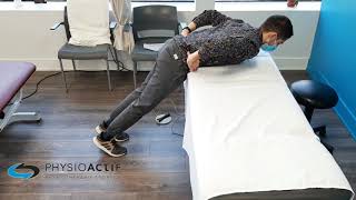
Make an appointmentWhat physiotherapy treatments are available?
Physiotherapy treatment aims to reduce irritation and improve hip muscle control. Studies show that 70% of patients can be successfully treated without surgery.
Initial assessment:Your physiotherapist will perform a complete assessment to identify the factors contributing to your condition:
- Your joint mobility
- The gliding of your nerves
- The quality of your movements
- Your muscle strength and stability
- Your movement patterns during your activities
Based on the assessment results, your physiotherapist will be able to:
- Mobilizing your hip to reduce pain and improve movement
- Provide you with exercises to strengthen your gluteal and hip-stabilizing muscles
- Teach you neuromuscular control exercises to improve the quality of your movements
- Help you temporarily modify activities that irritate your hip
- Give you advice for your posture and daily movements
A typical rehabilitation program lasts about 12 weeks, with 6 to 10 sessions with your physiotherapist. Studies show that improvements are maintained long-term, even after 5 years of follow-up.
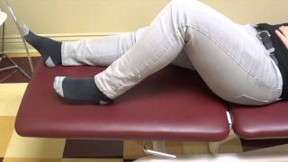
What to do at home?
Temporarily reduce movements that increase your pain and avoid hip compression positions. Here are some practical tips:
Recommended activities:- Swimming (all styles)
- Cycling or stationary bike (with a well-adjusted seat)
- Elliptical trainer
- Moderate walking on flat ground
- Standing with your weight shifted to one hip
- Sitting with your legs crossed
- Performing deep squats
- Forcing your knee towards your chest
If you are unable to sleep on your back, place a pillow between your thighs when lying on your side. This helps maintain your hip alignment.
Advice for athletes:- Incorporate walking breaks into your running sessions
- Temporarily reduce the intensity and volume of your training
- Do glute strengthening exercises regularly
- Avoid movements that reproduce your pain
If you don't see any improvement after 10 to 14 days of modifying your activities, consult a physiotherapist.
What is the recovery time?
Most people see significant improvement within 8 to 12 weeks with appropriate physiotherapy treatment. Continuing the exercises is important for long-term success.
Factors influencing healing time:- How long you've had symptoms (more recent = faster recovery)
- Your adherence to the recommended exercises
- Your Physical Activity Level
- The severity of your condition
Studies show that improvements gained with physiotherapy are maintained even after 5 years. The secret is to continue your strengthening exercises even after your symptoms disappear.
When to consider surgery?Surgery (hip arthroscopy) is only considered if conservative treatment has not worked after at least 2 months of consistent effort. The vast majority of people will not need surgery.
Sources
- Institut de kinésithérapie Paris. Femoroacetabular Impingement. Canadian study prevalence.
- Kuhns BD, et al. Femoroacetabular Impingement. StatPearls [Internet]. 2024.
- Source: Physioactif clinic.
- Cleveland Clinic. Hip Impingement (Femoroacetabular Impingement or FAI).
- Agricola R, et al. The Prevalence of Cam and Pincer Morphology and Its Association With Development of Hip Osteoarthritis. J Orthop Sports Phys Ther. 2018;48(4):230-238.
- Kemp JL, et al. Optimizing Conservative Treatment for Femoroacetabular Impingement Syndrome: A Scoping Review. Applied Sciences. 2025;15(5):2821.
- Femoroacetabular Impingement: Critical Analysis Review of Current Nonoperative Treatments. PMC. 2024.
- Griffin DR, et al. The FASHIoN trial: personalised hip therapy. Lancet. 2018.
- British Hip Society. Conservative Treatment for Femoro-Acetabular Impingement Syndrome. Guidelines 2024.
.jpg)
Other conditions
Hip osteoarthritis is the normal wear and tear of the hip joint. It is often said that osteoarthritis is the wear and tear of the cartilage between our bones. That is true, but it involves more than just the cartilage. Cartilage is a tissue that acts as a cushion between the surfaces of our bones and allows our joints to glide smoothly and move fluidly.
This is normal wear and tear of the knee joint. It’s often said that osteoarthritis is the wearing down of the cartilage between our bones. That’s true, but it’s more than just the cartilage. Cartilage is a tissue that acts as a cushion between the surfaces of our bones and allows our joints to glide smoothly and move fluidly.
It is an inflammation of the subacromial bursa in the shoulder joint.
A bursa is a small, thin sac filled with fluid that is found in many of the body's joints. This small sac acts as a cushion within the joint and lubricates the structures that are subject to increased friction.
It is a tissue that surrounds the shoulder and helps keep the shoulder bone in place within the joint. The capsule helps keep the joint stable.
Neck pain is a general term used to describe pain in the neck that has no specific cause, such as an accident or a sudden awkward movement. Neck pain is therefore synonymous with “my neck hurts, and nothing in particular happened.”
In both types of injury, pain is felt in the neck and then radiates into the arm, or vice versa.
Make an appointment now
We offer a three-pronged quality assurance approach: optimized treatment time, a second opinion from a physical therapist, and ongoing expertise to ensure effective care tailored to your needs.

Customer satisfaction is our top priority
At Physioactif, excellence guides everything we do, but our patients are the best ones to tell you about it. Take a look at their verified reviews to get a real sense of their experience.
Discover our physical therapy clinics
We have locations in several areas to better serve you.
Blainville
190 Bas-de-Sainte-Thérèse Road, Suite 110,
Blainville, Quebec
J7B 1A7
Located in Blainville, near Rosemère, the Physioactif clinic is easily accessible to residents of the area and the surrounding communities
Laval
3224 Jean-Béraud Ave., Suite 220, Laval,
QC H7T 2S4
Located in Chomedey, in the heart of Laval, the Physioactif clinic is easily accessible to people in the area
Montreal
8801 Lajeunesse Street,
Montreal,
QC H2M 1R8
Located in Ahuntsic, near Villeray, the Physioactif clinic is easily accessible to residents of both neighborhoods
Saint-Eustache
180 25th Avenue, Suite
201 Saint-Eustache
QC J7P 2V2
Located in Saint-Eustache, the Physioactif clinic is easily accessible to residents of the area and the surrounding communities
Vaudreuil
21 Cité-des-Jeunes Boulevard, Suite 240,
Vaudreuil-Dorion, Quebec
J7V 0N3
Located in Vaudreuil-Dorion, the Physioactif clinic is easily accessible to people in the area
Make an appointment now
