Achilles tendon tear
It is a partial or complete tear of the Achilles tendon fibers.
.png)
.png)
.png)




Achilles Tendon Tear: Symptoms, Treatment, and Recovery
Sudden calf pain during sports. A whipping sensation. The inability to point your foot downwards. These signs often suggest an Achilles tendon tear. This injury primarily affects active individuals between 30 and 50 years old.
Here's the good news: with appropriate treatment and well-managed rehabilitation, the vast majority of patients regain their previous activity level.
As physiotherapists specializing in musculoskeletal rehabilitation, we regularly support patients after this injury. Physiotherapy plays a central role in your recovery, whether it's after surgery or with conservative treatment.
What is an Achilles Tendon Tear?
An Achilles tendon tear is a partial or complete rupture of the largest tendon in the human body. This tendon connects the calf muscles to the heel. It allows you to walk, run, and jump.
Achilles Tendon Anatomy
The Achilles tendon is 12 to 15 cm long. An area located 2 to 6 cm above the heel receives less blood flow. Most ruptures occur in this vulnerable area.2
The Achilles tendon withstands enormous loads: when running and jumping, up to about ten times the body's weight. That is why gradually and carefully reintroducing weight-bearing after a rupture is essential.
The tendon is formed by the joining of the gastrocnemius and soleus muscles. These muscles make up the calf and allow for plantar flexion of the foot (pointing your foot downwards).
Types of Tears
| Type | Definition | Residual function |
|---|---|---|
| Partial tear | A partial tear affects only some of the tendon's fibers, so the tendon remains partially functional. A complete tear severs the tendon across its entire width. | Partially functional tendon |
| Complete tear | Complete separation of ends | Severely compromised function |
Who is Most at Risk?
Ruptures affect 18 to 37 people per 100,000 annually.2 Men are 5 to 10 times more affected than women.
Achilles tendon ruptures are much more common in men: 2 to 12 times more common than in women.
Most Achilles tendon ruptures occur during sports—between 60 and 75 percent of cases—often in sports such as basketball or soccer.
| Risk factor | Impact |
|---|---|
| Age 30-50 years | Peak incidence |
| Fluoroquinolones | 2-6 times increased risk |
| Corticosteroids | Tendon weakening |
| Pre-existing tendinopathy | Increased risk |
| Explosive sports | Basketball, tennis, badminton |
What are the Symptoms of an Achilles Tendon Tear?
Symptoms include sudden calf pain, a whipping sensation, difficulty pointing the foot downwards, and rapid swelling above the heel.
Signs of a Complete Tear
- Sudden, sharp pain
- Hearing or feeling a popping sound at the moment of injury suggests a tear. This popping sound is described as a blow to the back of the leg.
- Inability to point the foot downwards against resistance
- A palpable gap in the tendon (the 'gap')
- Significant difficulty or inability to walk normally
- Swelling and bruising that appear in the following hours
Signs of a partial tear
- Calf pain but partial ability to point the foot
- Weakness when walking or climbing stairs
- Localized tenderness when the tendon is touched
- Moderate swelling
How is an Achilles tendon tear diagnosed?
Diagnosis is based on a clinical examination, including the Thompson test, feeling for a gap in the tendon, and if necessary, an ultrasound or MRI to confirm the extent of the injury.
Your physiotherapist or doctor will perform several tests:
| Test | Method | Interpretation | Reliability |
|---|---|---|---|
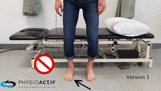
| Thompson Test | Calf compression while lying on stomach | No flexion = probable rupture | When you press on the calf and the foot does not move, this strongly suggests a complete tendon rupture. This is the basis of the Thompson test, which is used to detect a complete rupture. |
| Gap palpation | Checking for a defect in the tendon | Palpable gap = confirmed rupture | 73-89% |
| Matles Test | Foot observation with knee bent at 90° | Foot drop = rupture | 88% |
Ultrasound confirms the diagnosis with 95-100% sensitivity.3 MRI is reserved for cases where a partial rupture is suspected or for surgical planning.
The condition may be missed at first: about 25% of acute tears go unnoticed, since other muscles can still flex the foot. That is why a popping sound followed by pain warrants an examination, even if the foot can still move.
10 Quick Tips for Understanding Your Pain
The ones that have made the biggest difference in my patients' lives. 1 a day, 2 minutes.
Surgery or conservative treatment: which option to choose?
The choice depends on your age, activity level, and preferences. Surgery offers a lower re-rupture rate but carries risks. Conservative treatment with a functional protocol yields comparable results for many patients.
Comparing the two approaches
| Criterion | Surgery | Conservative |
|---|---|---|
| Re-rupture rate | 2-4% | 4-5% (modern protocol) |
| Risk of infection | 2-4% | None |
| Healing complications | Complications such as infection are more common when surgery is performed. A meta-analysis reports a complication rate of 4.9% after surgery, compared with 1.6% without surgery. | |
| Final strength recovered | Calf strength recovers well, but rarely to its full potential: a 10 to 30 percent deficit compared to the uninjured side is common. That is why strengthening exercises must be continued for a long time. | |
| Return to office work | 6-8 weeks | 8-10 weeks |
| Profile | Recommended option | Justification |
| Athlete, under 40 years old | Surgery | Optimal strength, lower risk of re-rupture |
| Large gap (over 5 cm) | Surgery | Difficult to bring together without intervention |
| Over 50 years old, sedentary | Conservative | Avoids surgical risks |
| Comorbidities (diabetes, etc.) | Conservative | Increased healing risks |
Modern functional protocols, involving early weight-bearing and rapid mobilization, have significantly improved the outcomes of conservative treatment.4
Today, non-surgical treatment combined with supervised rehabilitation yields re-rupture rates similar to those of surgery. You should discuss your options with your surgeon based on your individual circumstances.
New ruptures are rare: about 2.3% after surgery and 3.9% without surgery. With early functional rehabilitation, this difference disappears.
Surgery is not always the best option. For some people—including those with diabetes, nerve damage, excess weight, or a sedentary lifestyle—a cautious approach is recommended before undergoing surgery.
How does physiotherapy help you recover from an Achilles tendon tear?
Physical therapy plays a key role in recovery from an Achilles tendon rupture. It includes progressive mobilization, gradual eccentric strengthening, proprioception, and preparation for a return to activities.
Staying completely still is not the goal. Early, supervised movement of the tendon after repair promotes recovery and improves blood flow to the tendon.
The phases of rehabilitation
Our approach is based on the latest functional protocols. We promote early and safe weight-bearing:
| Phase | Duration | Goals | Treatments |
|---|---|---|---|
| 1. Protection | 0-2 weeks | Initial healing | Boot, gentle passive movements |
| 2. Mobilization | 2-6 weeks | Range of motion | Active ROM, partial weight-bearing |
| 3. Initial strengthening | 6-12 weeks | Basic strength | Light isometric and eccentric exercises |
| 4. Advanced Strengthening | 12-20 weeks | Functional Strength | Eccentric exercises, proprioception |
| 5. Return to Sport | 20+ weeks | Performance | Plyometrics, sport-specific exercises |
Techniques used in physiotherapy
- Ankle joint mobilization
- Soft tissue manual therapy
- Progressive eccentric strengthening program
- Proprioception and balance exercises
- Education on load management
Criteria for returning to sports
- Plantar flexion strength greater than 90% of the healthy side
- Ability to perform more than 25 single-leg calf raises
- Before allowing a return to sports, we look for near symmetry between the injured leg and the other leg during jump tests. The exact threshold depends on the type of test used.
- No pain during daily activities
How long does it take to recover from an Achilles tendon tear?
Recovery generally takes 4 to 6 months to resume normal daily activities and 6 to 12 months for a return to competitive sports. These timelines vary depending on the severity of the injury and the type of treatment.
Normal walking usually returns between 12.5 and 18 weeks after the injury. Early weight-bearing and rehabilitation help facilitate this recovery.
About 80% of people return to sports after an Achilles tendon rupture, although the timeline varies greatly from person to person.
Need professional advice?
Our physical therapists can assess your condition and provide you with a personalized treatment plan.
Make an appointment| Step | Approximate Timeline | What you can do |
|---|---|---|
| Walking without a boot | 6-8 weeks | Normal walking without limping |
| Return to office work | 6-10 weeks | Able to sit comfortably |
| Return to physical work | 12-16 weeks | Demanding physical tasks |
| Recreational sports | 4-6 months | Swimming, cycling, light jogging |
| Competitive sport | The return to sports is a process that takes several months to prepare for and is determined by strength and functional tests, not by a specific date on the calendar. Light activities resume well before competitive sports. | Full return based on tests |
Factors influencing your recovery
- Consistency with rehabilitation exercises
- Type of treatment chosen (slight advantage for surgery)
- Age and activity level before the injury
- Initial tendon quality
What exercises should you do to rehabilitate your Achilles tendon?
Rehabilitation progresses from passive mobility exercises to gentle stretching, then to isometric and eccentric strengthening, and finally to plyometric exercises. It is important to allow the tissue to heal properly.
Here are the exercises we prescribe to our patients based on their recovery phase:
Initial Phase (0-6 weeks)
- Ankle raises. Flexion and extension movements of the foot without bearing weight
- Alphabet with Your Feet. Draw the letters of the alphabet with your toes to improve mobility
- Isometric contractions. Gently push your foot against a fixed resistance without moving.
Intermediate Phase (6-12 weeks)
- Bilateral lifts. Stand on your tiptoes with both legs.
- Gentle stretches. Calf and soleus muscles against a wall
- Stationary bike. Light resistance, pain-free
- Progressive walking. Gradually increase the distance
Advanced Phase (12+ weeks)
- Single-leg lifts. Rise onto your toes using only the affected leg
- Off-center on the step. Slowly lower your heel below the level of the step
- Proprioceptive exercises. Balancing on one leg, unstable surfaces
- Introduction to Jogging. Start once you meet the strength requirements
Your physiotherapist will adjust this progression based on your recovery and personal goals.
How to prevent a new Achilles tendon rupture?
To prevent a recurrence, maintain a regular eccentric strengthening program, warm up adequately before physical activity, gradually increase your workout intensity, and consult a professional if tendon pain persists.
| Prevention Strategy | Practical Application | Frequency |
|---|---|---|
| Eccentric strengthening | Step-ups, slow descents | 2-3 times per week |
| Adequate warm-up | 10-15 minutes before activity | Each session |
| Gradual Progression | The load should be increased gradually, based on the tendon's tolerance rather than on a fixed percentage increase. | Ongoing |
| Monitor symptoms | Consult if pain persists | As needed |
With complete rehabilitation and these preventive measures, the recurrence rate is between 3 and 6%.5
When to consult a physiotherapist?
Consult a professional quickly if you experience the symptoms described in this article. Early diagnosis allows for appropriate treatment to begin without delay, optimizing your recovery.
You do not need to see a doctor before consulting a physiotherapist. If your condition requires surgery or medical advice, your physiotherapist will refer you to the right specialist.
Sources
- Józsa L, Kannus P. Human Tendons: Anatomy, Physiology, and Pathology. Champaign, IL: Human Kinetics; 1997.
- Park SH, Lee HS, Young KW, Seo SG. Treatment of Acute Achilles Tendon Rupture. Clin Orthop Surg. 2020;12(1):1-8. doi:10.4055/cios.2020.12.1.1
- Ochen Y, Beks RB, van Heijl M, et al. Operative treatment versus nonoperative treatment of Achilles tendon ruptures: systematic review and meta-analysis. BMJ. 2019;364:k5120. doi:10.1136/bmj.k5120
- Willits K, Amendola A, Bryant D, et al. Operative versus nonoperative treatment of acute Achilles tendon ruptures: a multicenter randomized trial using accelerated functional rehabilitation. J Bone Joint Surg Am. 2010;92(17):2767-75.
- Barfod KW, Bencke J, Lauridsen HB, et al. Nonoperative dynamic treatment of acute Achilles tendon rupture: the influence of early weight-bearing on clinical outcome. J Bone Joint Surg Am. 2014;96(18):1497-503.
.jpg)
.jpg)
Other conditions

Hip osteoarthritis is the normal wear and tear of the hip joint. It is often said that osteoarthritis is the wear and tear of the cartilage between our bones. That is true, but it involves more than just the cartilage. Cartilage is a tissue that acts as a cushion between the surfaces of our bones and allows our joints to glide smoothly and move fluidly.
This is normal wear and tear of the knee joint. It’s often said that osteoarthritis is the wearing down of the cartilage between our bones. That’s true, but it’s more than just the cartilage. Cartilage is a tissue that acts as a cushion between the surfaces of our bones and allows our joints to glide smoothly and move fluidly.
It is an inflammation of the subacromial bursa in the shoulder joint.
A bursa is a small, thin sac filled with fluid that is found in many of the body's joints. This small sac acts as a cushion within the joint and lubricates the structures that are subject to increased friction.
It is a tissue that surrounds the shoulder and helps keep the shoulder bone in place within the joint. The capsule helps keep the joint stable.
Neck pain is a general term used to describe pain in the neck that has no specific cause, such as an accident or a sudden awkward movement. Neck pain is therefore synonymous with “my neck hurts, and nothing in particular happened.”
In both types of injury, pain is felt in the neck and then radiates into the arm, or vice versa.
Make an appointment now
We offer a three-pronged quality assurance approach: optimized treatment time, a second opinion from a physical therapist, and ongoing expertise to ensure effective care tailored to your needs.
Customer satisfaction is our top priority
At Physioactif, excellence guides everything we do, but our patients are the best ones to tell you about it. Take a look at their verified reviews to get a real sense of their experience.









Discover our physical therapy clinics
We have locations in several areas to better serve you.
Blainville
190 Bas-de-Sainte-Thérèse Road, Suite 110,
Blainville, Quebec
J7B 1A7
Located in Blainville, near Rosemère, the Physioactif clinic is easily accessible to residents of the area and the surrounding communities
Boucherville
690 Rue de Montbrun, Suite S,
Boucherville, Quebec
J4B 8H2
Located in Boucherville, the Physioactif clinic is easily accessible to people in the area
Laval
3224 Jean-Béraud Ave., Suite 220, Laval,
QC H7T 2S4
Located in Chomedey, in the heart of Laval, the Physioactif clinic is easily accessible to people in the area
Montreal
8801 Lajeunesse Street,
Montreal,
QC H2M 1R8
Located in Ahuntsic, near Villeray, the Physioactif clinic is easily accessible to residents of both neighborhoods
Saint-Eustache
180 25th Avenue, Suite
201 Saint-Eustache
QC J7P 2V2
Located in Saint-Eustache, the Physioactif clinic is easily accessible to residents of the area and the surrounding communities
Vaudreuil
21 Cité-des-Jeunes Boulevard, Suite 240,
Vaudreuil-Dorion, Quebec
J7V 0N3
Located in Vaudreuil-Dorion, the Physioactif clinic is easily accessible to people in the area
Make an appointment now