Lumbo-sciatica
.png)
.png)
.png)


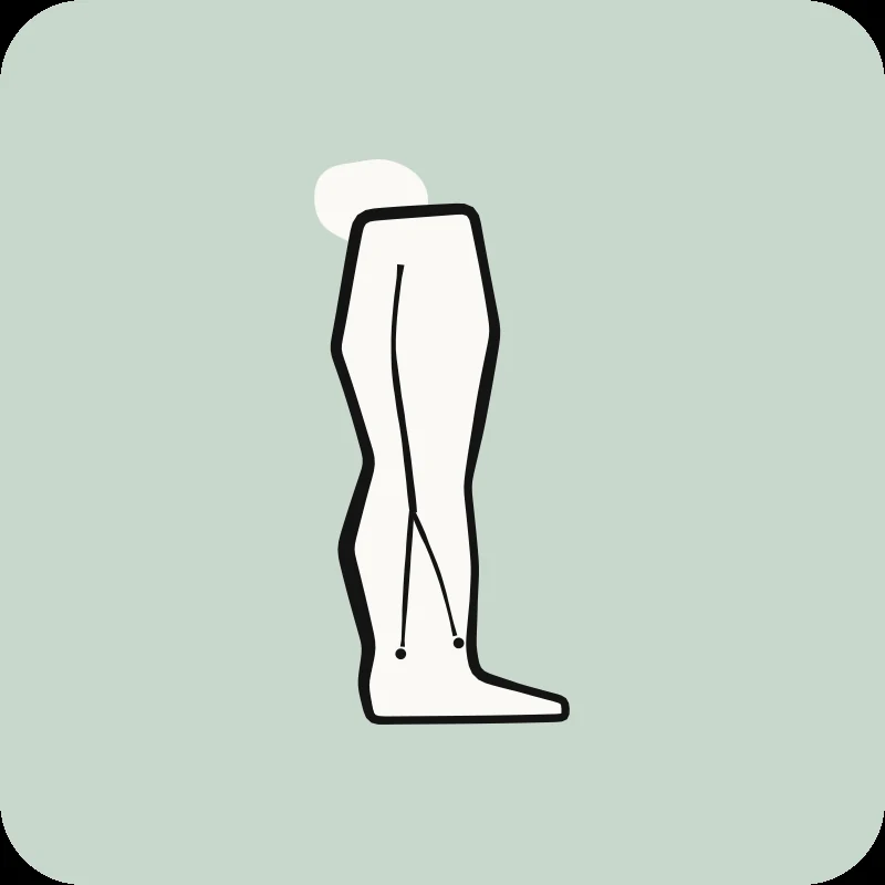


The pain starts in your lower back and travels down your leg, sometimes all the way to your foot, feeling like a burning sensation, electric shocks, or numbness that follows a specific path. You wonder if it’s serious, if you’ll need surgery, or if it will go away. This worry is normal. Sciatica affects 10 to 40% of the population at some point in their lives.¹
Good news: In 80 to 90% of cases, you can recover without surgery.³ Physiotherapy offers effective treatments that address the cause of nerve compression. What science teaches us about sciatica:- Sciatica is a symptom, not a disease. Understanding its cause allows for more effective treatment.
- A herniated disc causes 90% of acute cases, and most resolve naturally.
- The position that provides relief varies depending on the cause (sitting vs. standing, flexion vs. extension).
- Symptoms that extend below the knee indicate true nerve compression.
This guide explores the causes of your sciatica and how physiotherapy can effectively relieve your pain. To understand the broader context, consult our complete guide to back pain.
What is sciatica and which nerve does it affect?
Sciatica refers to pain that follows the path of the sciatic nerve. It starts in your lower back, runs through your buttock, and down your leg. This nerve can become irritated or compressed, causing pain, numbness, and muscle weakness along its path down to just below the knee.
Sciatica is a symptom, not a diagnosis in itself.² The term describes pain that follows the anatomical path of the sciatic nerve. This nerve is formed by five nerve roots (L4, L5, S1, S2, and S3). They converge in your lower back and pelvis.⁴ This nerve is about as wide as your thumb at its origin, making it the largest single nerve in the human body.² After emerging from the spine, the sciatic nerve travels deep into your buttock. It passes under the piriformis muscle (a small muscle in the buttock) and then runs down the back of your thigh. There, it branches into smaller nerves that supply the lower leg and foot.²
True sciatica involves actual compression or irritation of the sciatic nerve roots or the nerve itself. It produces specific neurological symptoms that follow precise patterns.⁵ It differs from referred pain, which can radiate down the leg without following the exact path of the sciatic nerve or presenting associated neurological signs such as changes in reflexes or loss of sensation.⁵ This distinction is important. True sciatica indicates nerve damage that requires specific treatments. Referred pain generally responds to other interventions that target the source tissue.
Each year, 1 to 5% of people develop sciatica.¹ Men are slightly more affected than women. The peak incidence occurs in people in their 40s and 50s.¹ Certain jobs increase the risk, such as heavy lifting, prolonged sitting, or repeated twisting movements.⁶
The majority of sciatica cases stem from lower back problems. Discover our guide on lower back pain to understand the different conditions that can cause sciatica.
What causes sciatic pain?
Sciatica results from compression or irritation of the sciatic nerve. In 90% of acute cases, it's caused by a lumbar disc herniation. Other causes include spinal stenosis (narrowed canal), piriformis syndrome (overly tight buttock muscle), or spondylolisthesis (a vertebra that slips forward). Less common causes include tumors, infections, or pregnancy-related pressure on nerve roots.
A lumbar disc herniation causes the vast majority of acute cases of sciatica. It occurs when the soft center of the disc (nucleus pulposus) protrudes through its outer layer (annulus fibrosus). It then compresses the adjacent nerve roots.⁷ This compression creates two problems. First, direct mechanical pressure. Second, chemical irritation caused by inflammatory mediators (irritating substances) released by the disc material.⁸ The L4-L5 and L5-S1 discs are most commonly affected because these segments bear the heaviest mechanical loads during your daily activities.⁷
A herniated disc is a common cause of acute sciatica. A herniated disc may resolve on its own over time, even without surgery. See our comprehensive guide to lumbar disc herniation for information on the mechanisms of herniation, recovery times, and treatment approaches.
Reassuring news: In people who have no back pain, imaging often reveals abnormalities. In one study, 28% had a herniated disc and 52% had disc bulging. An abnormal image, therefore, does not always explain the pain.
When monitored over several years, most herniated discs remain stable or improve. About 65% do not worsen, and only 12.5% get worse.
Spinal stenosis is the second most common cause. It involves a narrowing of the spinal canal (the bony tunnel) or the openings where nerve roots exit the spine.⁹ This narrowing can result from age-related degenerative changes. These include disc height loss, osteoarthritis of the facet joints (small joints in the back), and thickening of the ligamentum flavum (a thick ligament in the back).⁹ Unlike disc herniation, stenosis generally develops gradually. It affects older adults. Its symptoms worsen when standing or walking and improve when sitting or leaning forward.⁹
Are you over 60? Does your sciatica worsen when standing or walking but improve when sitting or leaning forward (like when pushing a grocery cart)? Spinal stenosis could be your primary cause. The treatment approach differs from that for disc herniation. Consult our complete guide to spinal stenosis for tailored management strategies.
Piroiformis syndrome is a peripheral cause of sciatica, in which the piriformis muscle in your buttock compresses the sciatic nerve.¹⁰ This muscle lies directly over the sciatic nerve. A muscle spasm (involuntary contraction) or anatomical variations can cause nerve irritation. This mimics spinal sciatica.¹⁰ Spondylolisthesis, in which one vertebra slips forward over another, can also compress the nerve roots. This produces symptoms of sciatica.¹¹
Less common causes include spinal tumors, infections like epidural abscess (an infection in the back), and pregnancy-related nerve compression.¹² Risk factors include obesity, a sedentary lifestyle, physically demanding jobs, diabetes, and smoking. Smoking negatively impacts disc nutrition and healing.⁶
What are the characteristic symptoms of sciatica?
If you’re experiencing pain that radiates down your leg, you’re not alone: millions of people go through the same thing every year. Symptoms of sciatica include sharp, burning, or electric-like pain. It starts in the lower back, travels through the buttock, and down one leg, often extending below the knee. You may also experience numbness, tingling, or muscle weakness. The pain worsens when sitting, leaning forward, or coughing.
The hallmark of sciatica is unilateral leg pain that extends below the knee. It is typically more severe than any accompanying back pain.⁵ Patients describe the pain quality as sharp, shooting, burning, or electric-shock-like. These sensations follow the distribution of the sciatic nerve.¹³ The pain pattern provides diagnostic clues. Compression of the L5 nerve root produces pain that radiates down the back and side of the thigh, continuing to the side of the calf and the top of the foot. S1 compression causes pain in the back of the thigh, calf, and side of the foot.⁵
Sensory symptoms include numbness, tingling, or altered sensation in the affected leg area.⁵ These sensory changes follow specific patterns, corresponding to the level of the compressed nerve root. Motor symptoms can also develop, including weakness in specific muscle groups. L5 compression affects the upward movement of the ankle and big toe, leading to what is called foot drop. S1 compression weakens the downward movement of the ankle and also affects big toe flexion.¹⁴ Deep reflexes may decrease. L5 compression does not affect any specific reflex, while S1 compression reduces the Achilles reflex (at the ankle).¹⁴
Numbness is the most commonly reported symptom of sciatica caused by a herniated disc: about 94% of people describe experiencing it. It is a common manifestation of nerve irritation, not a sign of severity.
The pain typically worsens with activities that increase pressure on the disc. These include sitting, bending forward, coughing, or sneezing.¹⁵ Many patients report relief when lying down or standing, but individual responses vary depending on the specific condition and directional preference.¹⁵ Sciatica usually occurs on one side (one leg). However, severe central disc herniations or spinal stenosis can cause bilateral symptoms, affecting both legs simultaneously.⁵
Red flags requiring immediate medical attention:¹⁶These serious symptoms are rare. However, it is important to be aware of them. Go to the emergency room immediately if you have:
- Progressive muscle weakness that worsens over several days
- Loss of bladder or bowel control (cauda equina syndrome, severe nerve compression).
- Numbness in the genital or anal region
- Symptoms in both legs simultaneously
- Severe and progressive neurological deficits
These symptoms suggest an urgent compression of the spinal cord or cauda equina. This requires an emergency evaluation. Sometimes, surgical decompression within 48 hours is required.¹⁶
10 Quick Tips for Understanding Your Pain
The ones that have made the biggest difference in my patients' lives. 1 a day, 2 minutes.
How do healthcare professionals diagnose sciatica?
Diagnosing sciatica involves a clinical examination with specific tests. This includes the straight leg raise test, a neurological assessment of reflexes and muscle strength, and a review of your medical history. Rest assured: clinical findings usually provide enough information for a diagnosis. MRI scans are not necessary in most cases and are reserved for severe situations or when surgery is being considered.
The clinical examination begins with a detailed history. Your physiotherapist will characterize the pain: its location, quality, path, and what makes it worse or better.¹⁷ We also assess your posture, gait (how you walk), and spinal mobility. We note any antalgic posture (a position to avoid pain) or limitation of movement.¹⁷ The neurological examination systematically tests motor strength, sensory function, and deep reflexes. This identifies the specific nerve root level affected.¹⁴
What clinical tests identify sciatic nerve involvement?
Your physiotherapist uses specific tests to identify sciatic nerve compression. The table below shows the most common clinical tests:
Test Name
How it's performed
Positive Sign
Sensitivity
Specificity
Straight leg raise¹⁸Your therapist raises your straight leg while you are lying on your back
Pain that travels below the knee between 30-70° of hip flexion
91% (detects most cases)
26% (low)
Crossed leg raise¹⁸Raising the unaffected leg reproduces pain in the affected leg
Pain in the affected leg
29% (low)
88% (high)
Slump test¹⁹Combines spinal flexion, hip flexion, and knee extension
Pain with neck flexion, relief with neck extension
Moderate
Moderate
Strength Test¹⁴Tests ankle and toe movement
Weakness: L5 (foot drop), S1 (pushing ankle down)
Variable
Variable
Reflex test¹⁴Taps the knee and Achilles tendon
Reduced reflexes: L4 (knee), S1 (Achilles)
Moderate
Moderate
Sensory test¹⁴Maps areas of numbness
Numbness follows a specific nerve root pattern
Variable
Moderate
The straight leg raise test is the most useful. It identifies 91% of disc herniation cases. However, it can also be positive in other conditions, meaning it has low specificity. The crossed straight leg raise is less common but more specific for disc-related issues.
Routine neurological tests alone are not sufficient to confirm a herniated disc. Sensory testing detects herniated discs in about 40% of cases, and reflex testing in about 29%.
When is an MRI necessary to diagnose sciatica?
Imaging studies, especially MRI, are not routinely required for the initial management of sciatica. A clinical examination provides adequate diagnostic information in most cases.²⁰ Clinical guidelines recommend against early imaging for patients without "red flags" (warning signs). MRI results frequently show abnormalities in individuals without symptoms, and these findings do not necessarily correlate with symptoms or predict recovery.²⁰ Approximately 30 to 40% of asymptomatic individuals show disc herniations on MRI, demonstrating that imaging results alone do not determine the need for treatment.²¹
For sciatica, the herniation doesn't have to disappear for symptoms to improve. In a follow-up study, about 84% of people made a full recovery in less than a year, regardless of whether the herniation was still visible on an MRI.
An MRI is indicated when severe or progressive neurological deficits develop, when symptoms persist despite several weeks of properly administered conservative treatment, when surgery is being considered, or when red flags suggest a serious underlying condition. This includes a tumor, an infection, or cauda equina syndrome.²⁰
Differential diagnosis helps distinguish sciatica from other conditions such as piriformis syndrome, hip problems, peripheral neuropathy, and vascular claudication.²² An accurate diagnosis ensures appropriate treatment that targets the true source of pain. Refer to the section on differences with other conditions further down in this article for the distinctive characteristics of each condition.
How does physiotherapy effectively treat sciatica?
Physical therapy treatment combines nerve mobilization techniques, specific exercises based on your directional preference, manual therapy, activation of stabilizing muscles, and progressive strengthening. This reduces nerve irritation and restores function. This treatment addresses both the source of nerve compression and secondary compensations. Conservative treatment, including evidence-based physical therapy, allows some people to avoid or delay surgery.
Science-based physiotherapy for sciatica uses multimodal treatment approaches tailored to your specific symptoms and underlying cause.²³ The treatment strategy evolves through distinct phases. Acute phase management focuses on reducing pain and nerve irritation. The subacute phase emphasizes restoring mobility and correcting movement patterns. The chronic phase prioritizes strengthening, functional restoration, and preventing future recurrences.²³
Science demonstrates that nerve gliding techniques significantly reduce irritation of the sciatic nerve. Discover our approach to nerve mobilization to restore nerve gliding and reduce mechanical sensitivity.
Neural mobilization techniques, also known as nerve glide exercises, promote nerve movement through the surrounding tissues. This reduces adhesions and mechanical sensitivity.²⁴ These gentle techniques involve alternating positions. They lengthen and shorten the nerve sheath, which restores normal gliding and reduces chemical irritation.²⁴ Research shows that neural mobilization significantly reduces pain. It improves function in patients with sciatica when combined with other interventions.²⁴
Research supports the use of specific directional movements to treat sciatica related to herniated discs. One study shows that approximately 72% of patients exhibit a directional preference. Moving in this preferred direction leads to better outcomes than moving against it. Our McKenzie approach is a science-based method that identifies each patient’s directional preference through a systematic evaluation.²⁵ Most patients with sciatica show an extension preference. This means that extension movements toward the back centralize or reduce pain in the leg.²⁵ However, some patients demonstrate preferences for flexion or lateral movement. This underscores the importance of individualized assessment rather than protocol-based treatment.²⁵
Manual therapy techniques include spinal mobilization, manipulation, soft-tissue therapy to release muscle guarding (protective muscle tension), and joint mobilization to restore normal spinal mechanics.²⁶ Scientific evidence supports the use of manual therapy in combination with exercise, an approach that is more effective than either intervention alone. The greatest benefits are seen in the subacute and chronic phases.²⁶ Your physical therapist selects specific manual techniques based on the results of your evaluation, the phase of treatment, and your individual response.²⁶
Progressive therapeutic exercise forms the foundation of successful sciatica treatment.²³ Early-phase exercises focus on gentle movements that do not aggravate symptoms. These include directional preference exercises, pelvic tilts, and supported positions. As symptoms improve, progressing to deep core strengthening becomes essential. Scientific evidence shows that patients who maintain their lumbar strength can reduce their risk of recurrence. Our exercises for the stabilizing muscles progress to core stabilization training, hip strengthening, flexibility exercises for tight muscle groups, and functional movement training.²³ A systematic review found that ourmuscle-strengthening and endurance exercise programs significantly improve pain and function compared to usual care or no treatment for chronic sciatica.²⁷
What is the expected recovery time for sciatica?
Sciatica often improves gradually with appropriate treatment. Many people notice an improvement within the first few weeks. Acute episodes often show a marked improvement within the first few weeks. More persistent cases may take longer to resolve. Complete resolution depends on the underlying cause, the severity of the condition, and your adherence to treatment.
The natural course of sciatica is generally favorable. Many cases show spontaneous improvement over time.²⁸ Acute sciatica often improves within the first few weeks of treatment. This occurs without any specific intervention. However, the rate of improvement increases substantially with appropriate physical therapy.²⁸ Acute sciatica (symptoms lasting less than 6 weeks) typically responds more quickly than chronic sciatica (symptoms lasting more than 12 weeks). Acute cases often show dramatic improvement within 2 to 4 weeks after the start of treatment.²⁹
As sciatica caused by a herniated disc improves, the pain often shifts from the leg to the lower back, and the symptoms as a whole become less bothersome over time. Seeing the pain move back up toward the back is actually a good sign.
Several factors influence the speed and completeness of recovery.³⁰
Factors that help you recover faster (positive signs):- Younger age
- Shorter delay before starting treatment
- Pain that centralizes (moves from the leg towards the spine)
- First episode of sciatica
- Active participation in prescribed exercises
- Older Age
- Severe disability at the onset
- Depression or anxiety
- Heavy physical work demands
- Involvement in workers' compensation or litigation
- Widespread pain beyond the typical sciatica pattern
Early intervention with physiotherapy speeds up recovery and prevents the condition from becoming chronic pain.³⁹ Patients who start treatment within the first few weeks achieve better results than those who delay.³⁹ This early window allows for addressing movement patterns, educating patients about their condition, and establishing effective self-management strategies.³⁹
Even chronic sciatica cases respond well to appropriate treatment. However, recovery times are longer compared to acute presentations.²⁹ Patients with symptoms lasting beyond 12 weeks often require more intensive rehabilitation. This addresses secondary issues such as deconditioning, fear-avoidance behaviors, and altered movement patterns, which develop during the acute phase.²⁹ With comprehensive physiotherapy addressing these factors, most patients with chronic sciatica achieve significant functional improvement within 8 to 12 weeks. However, complete resolution of symptoms may take longer.²⁹
Need professional advice?
Our physical therapists can assess your condition and provide you with a personalized treatment plan.
Make an appointmentWhat positions and movements help relieve sciatica pain?
Relieving positions include lying on your back with bent knees, lying on your side with a pillow between your knees, and avoiding prolonged sitting. Gentle walking, specific directional exercises determined by evaluation, and nerve mobilization movements can provide significant pain reduction when performed correctly.
Postural adjustments are an essential part of self-management for sciatica. Certain postures reduce mechanical compression and chemical irritation of the sciatic nerve.³² Lying on your back with your knees bent and your feet flat reduces lumbar lordosis (the curve in the lower back). This reduces pressure on the disc compared to standing or sitting.³² Many patients find additional relief by placing pillows under their knees. This helps maintain this position comfortably while resting or sleeping.³² Lying on your side with a pillow between your knees maintains neutral spinal alignment. This prevents hip adduction (the legs moving toward each other).
Sitting for long periods typically worsens sciatica. It increases pressure on the disc by maintaining sustained spinal flexion, which can worsen the symptoms of a posterior disc herniation.³³ If sitting is necessary, providing lumbar support with a cushion or a rolled-up towel can help. Keeping the hips and knees at about a 90-degree angle is also helpful. Taking frequent breaks to stand up every 20 to 30 minutes helps minimize symptom flare-ups.³³ Some patients benefit from a slight anterior pelvic tilt while sitting. This creates a slight lumbar extension, which reduces posterior disc pressure.³³
Movement-based pain relief strategies depend on your individual directional preference, which is identified during the evaluation.²⁵ Patients with an extension preference typically experience pain relief with backward extension movements, such as push-ups in the prone position (cobra pose) or standing extension exercises.²⁵ These individuals should avoid activities involving prolonged flexion. This includes leaning forward, lifting with a rounded back, or slouching while seated.²⁵ Conversely, the minority of patients with a flexion preference find relief with forward-bending movements. They should minimize extension activities.²⁵
Gentle walking often provides relief. It promotes circulation, reduces muscle tension, and creates gentle, repetitive movements that facilitate nerve gliding.³⁴ Walking keeps the spine in a neutral position or slight extension. This avoids prolonged static postures. This makes it an ideal activity for most patients with sciatica during the acute phase.³⁴ Nerve mobilization exercises, when taught by your physical therapist and performed within a comfortable range of motion, promote the movement of the sciatic nerve through the surrounding tissues. This reduces mechanical sensitivity.²⁴
Activities to temporarily avoid during acute sciatica include heavy lifting, bending forward with rotation, sitting for long periods without breaks, high-impact activities such as running or jumping, and any movement that consistently triggers or worsens symptoms in the leg.³⁵ The individualized nature of sciatica symptoms means that the specific positions and movements that provide relief vary from patient to patient. This underscores the importance of a professional evaluation. It allows for the identification of your specific movement preferences AND the creation of a personalized management strategy.
When should surgery be considered for sciatica?
Here's some good news: the vast majority of people with sciatica will never need surgery. It is rare for sciatica to require surgery. Surgery is primarily considered in cases of progressive muscle weakness, loss of bowel or bladder control, or when conservative treatment has not worked after 6 to 12 weeks despite severe functional limitations.
Surgery for sciatica is reserved for specific indications, such as when conservative treatment proves insufficient or when urgent decompression is medically necessary.³⁶ The most urgent indication for surgery is cauda equina syndrome. It is characterized by progressive bilateral weakness in the legs, saddle numbness (genital and anal area), and bowel or bladder dysfunction. It requires emergency decompression within 48 hours to prevent permanent neurological damage.¹⁶ This rare complication occurs in fewer than 2% of cases of herniated discs. However, it represents a true surgical emergency.¹⁶
Progressive motor weakness despite conservative treatment represents another strong surgical indication. Prolonged nerve compression can lead to permanent muscle denervation if not addressed.³⁷ When muscle weakness worsens over days to weeks despite appropriate physiotherapy, or when significant weakness (muscle grade 3/5 or less) persists beyond 4 to 6 weeks, surgical consultation is appropriate.³⁷ The timing of intervention for motor weakness involves balancing the risk of permanent nerve damage against the high probability of natural resolution with conservative care.³⁷
For patients without progressive weakness or cauda equina syndrome, clinical guidelines recommend a 6- to 12-week trial of conservative treatment before considering surgery.³⁸ The decision to proceed with surgery after this period depends on the severity of pain, functional limitations, and patient preferences, rather than on an absolute failure of treatment.³⁸ The landmark SPORT (Spine Patient Outcomes Research Trial) study found that although surgical patients experienced faster initial improvement, long-term outcomes (2 to 4 years) showed similar pain and function scores between the surgical and conservative treatment groups.³⁹ This research demonstrates that surgery accelerates recovery. However, it does not necessarily improve final outcomes compared to well-structured physical therapy programs.³⁹
Surgical options for sciatica include microdiscectomy (removing the herniated portion of the disc compressing the nerve), laminectomy (removing bone to widen the spinal canal in cases of stenosis), or laminotomy (creating space for compressed nerve roots).⁴⁰ Minimally invasive techniques have reduced surgical complications and recovery times compared to traditional open procedures.⁴⁰ In carefully selected candidates, surgery may provide faster short-term relief. In the long term, the outcomes of surgery and conservative treatment tend to converge. However, long-term outcomes depend on factors including age, general health, smoking status, and participation in post-surgical rehabilitation.⁴⁰
Post-surgical rehabilitation with physiotherapy is essential for optimal outcomes and recurrence prevention.⁴¹ Surgery addresses mechanical compression; however, it does not correct the biomechanical factors, movement patterns, or muscle imbalances that contributed to the initial problem.⁴¹ Comprehensive post-operative physiotherapy addressing these factors significantly reduces the risk of recurrence and improves long-term functional outcomes.⁴¹
If you have undergone surgery for sciatica or are considering surgery, understanding how physical therapy supports postoperative recovery is crucial for optimal results. Post-surgical rehabilitation follows specific protocols. These differ from conservative treatment for sciatica. After surgery, a structured rehabilitation program helps you gradually resume your activities. Check out our information on lower back pain to learn more about the phases of rehabilitation, expected recovery milestones, and how physical therapy helps prevent complications.
How can you prevent sciatica from recurring?
Once your pain has improved, you have the power to significantly reduce the risk of it returning. Prevention involves maintaining adequate core strength, practicing proper body mechanics, regularly stretching hip and back muscles, adopting an ergonomic workplace setup, and maintaining a healthy weight. Addressing underlying biomechanical issues identified during physiotherapy significantly reduces the risk of recurrence.
Core stabilization is the foundation of sciatica prevention. Adequate strength and endurance in the core muscles protect the spine during daily activities. This reduces the forces on the intervertebral discs.⁴² The core musculature includes not only the abdominal muscles but also the multifidus, the transverse abdominis, the pelvic floor, and the diaphragm. They work together to create spinal stability.⁴² Research shows that individuals with a history of sciatica who maintain core exercise programs experience significantly lower recurrence rates compared to those who stop exercising once their symptoms resolve.⁴³ Your physical therapist designs progressive core-strengthening programs and helps restore functional movement patterns.⁴²
Proper body mechanics during lifting, bending, and daily activities minimize abnormal spinal loads. This could trigger a recurrence.⁴⁴ Key principles include maintaining neutral spinal alignment during lifts, squatting to lift objects off the ground rather than bending over with a rounded back, keeping heavy objects close to your body, avoiding twisting while holding loads, and breaking large loads into smaller carries.⁴⁴ Your physical therapist teaches these movement patterns through education and practice. This ensures that proper technique becomes second nature during daily activities.⁴⁴
Maintaining flexibility and mobility prevents muscle tension and joint restrictions that alter spinal mechanics and increase injury risk.⁴⁵ Regular stretching targeting hip flexors, hamstrings, piriformis, and lumbar extensors maintains optimal length-tension relationships, allowing for proper movement patterns.⁴⁵ Decreased hamstring flexibility, for example, increases posterior pelvic tilt during forward bending, placing excessive stress on the lumbar discs.⁴⁵ Incorporating daily stretching routines and activities like yoga or tai chi promotes flexibility and body awareness, providing significant protective benefits.⁴⁵
Ergonomic workplace modifications reduce cumulative postural stress that contributes to sciatica development.⁴⁶ For office workers, proper workstation setup includes a chair height allowing feet flat on the floor with knees and hips at 90 degrees, a monitor positioned at eye level to prevent neck flexion, lumbar support maintaining the spine's natural curves, and keyboard and mouse placement preventing shoulder elevation or excessive reaching.⁴⁶ Regular position changes, standing desk options, and micro-breaks every 30 minutes reduce sustained load and promote circulation.⁴⁶
Weight management impacts sciatica risk. Excess body weight increases mechanical loads on the lumbar discs and nerve roots.⁴⁷ Each kilogram of excess weight creates several times that force through the spine during daily activities.⁴⁷ Furthermore, obesity correlates with increased systemic inflammation and metabolic factors, which negatively impacts disc health and healing capacity.⁴⁷ Achieving and maintaining a healthy weight through balanced nutrition and regular physical activity provides significant protective effects against sciatica recurrence.⁴⁷
Smoking is a modifiable risk factor for disc degeneration and herniated discs in the lower back. Quitting smoking is one of the steps you can take to help protect your discs.
Addressing the underlying biomechanical abnormalities identified during the physical therapy evaluation is perhaps the most important preventive strategy.⁴⁸ This may include movement pattern dysfunctions requiring retraining of motor control, muscle imbalances requiring specific strengthening, or joint restrictions requiring ongoing mobility work.⁴⁸ Systematically correcting these contributing factors removes the mechanical stresses that triggered your initial episode of sciatica. This substantially reduces the likelihood of recurrence.⁴⁸
Prevention requires a personalized approach based on a thorough assessment of your specific biomechanical factors. Your physical therapist designs long-term prevention programs that address your unique risk factors and movement patterns. People who complete their rehabilitation and follow ergonomic recommendations can reduce their risk of recurrence. Explore our comprehensive information on lower back pain to discover science-based prevention strategies, maintenance exercise programs, and ongoing management approaches for all back pain conditions, including sciatica.
What distinguishes sciatica from other leg pain conditions?
Sciatica follows the path of the sciatic nerve, causing pain below the knee, unlike hip osteoarthritis or muscle tears, which remain localized. Vascular claudication (circulatory problems) improves with rest regardless of position. With sciatica, positioning matters. An accurate diagnosis ensures treatment tailored to your specific condition.
Sciatica can be confused with other leg pain conditions. The table below shows the key differences:
| Condition | Pain location | Key Differentiator | Clinical Test | Pattern |
|---|---|---|---|---|
| True sciatica⁵ | Lower back → buttock → below the knee | Pain follows sciatic nerve, neurological signs present | Positive SLR (Straight Leg Raise), reflex changes | Usually one leg |
| Hip Disorders⁴⁹ | Groin, side of hip, front of thigh | Worse with weight-bearing, better with rest | Positive FABER/FADIR tests | Localized to the hip |
| Vascular claudication⁵⁰ | Both calves equally | Better with rest (any position), predictable with walking distance | Diminished pulses, ankle-brachial index | Both legs symmetrical |
| Peripheral neuropathy⁵¹ | Both feet and lower legs | Glove-and-stocking pattern, gradual onset over months | Symmetrical sensory loss, intact reflexes | Both legs, starts in the feet |
| Piriformis syndrome¹⁰ | Buttock → back of thigh | Rarely goes below the knee, worse when sitting on hard surfaces | Positive FAIR test, no positive SLR | Dominant buttock |
| Referred pain (facet joint/sacroiliac joint)⁵² | Buttock and thigh only | Stops above the knee, no neurological signs | Facet/SI provocation tests | Local to the joint |
The key diagnostic feature of true sciatica is pain that extends below the knee. It follows the path of the sciatic nerve. It is combined with neurological signs such as changes in reflexes or numbness patterns. Other conditions remain more localized or show different patterns.
An accurate diagnosis is essential because the treatment approach varies significantly depending on the underlying cause. Treatment varies greatly depending on the underlying cause. An inaccurate initial diagnosis can delay recovery. See our guide on back pain for comprehensive information on understanding the different types of pain and their mechanisms.
Ready to find relief from your sciatic nerve pain?
Our physiotherapists at Physioactif provide a comprehensive sciatica evaluation and science-based treatment. We develop personalized programs that relieve pain, restore function, and prevent recurrence.
Don't let sciatic pain limit your activities. Contact Physioactif to schedule your evaluation. Our team will identify the cause of your sciatica and develop a treatment plan tailored to your goals. Take the first step towards recovery by booking your appointment now.
References
- Koes BW, van Tulder MW, Peul WC. Diagnosis and treatment of sciatica. BMJ. 2007;334(7607):1313-1317.
- Davis D, Maini K, Vasudevan A. Sciatica. In: StatPearls [Internet]. Treasure Island (FL): StatPearls Publishing; 2024 [cited 2025 Jan 10]. Available from: https://www.ncbi.nlm.nih.gov/books/NBK507908/
- Valat JP, Genevay S, Marty M, Rozenberg S, Koes B. Sciatica. Best Pract Res Clin Rheumatol. 2010;24(2):241-252.
- Giuffre BA, Jeanmonod R. Anatomy, Sciatic Nerve. In: StatPearls [Internet]. Treasure Island (FL): StatPearls Publishing; 2024 [cited 2025 Jan 10]. Available from: https://www.ncbi.nlm.nih.gov/books/NBK482431/
- Ropper AH, Zafonte RD. Sciatica. N Engl J Med. 2015;372(13):1240-1248.
- Shiri R, Karppinen J, Leino-Arjas P, Solovieva S, Viikari-Juntura E. The association between obesity and low back pain: a meta-analysis. Am J Epidemiol. 2010;171(1):135-154.
- Jordan J, Konstantinou K, O'Dowd J. Herniated lumbar disc. BMJ Clin Evid. 2011;2011:1118.
- Olmarker K, Rydevik B. Pathophysiology of sciatica. Orthop Clin North Am. 1991;22(2):223-234.
- Lurie J, Tomkins-Lane C. Management of lumbar spinal stenosis. BMJ. 2016;352:h6234.
- Hopayian K, Song F, Riera R, Sambandan S. The clinical features of the piriformis syndrome: a systematic review. Eur Spine J. 2010;19(12):2095-2109.
- Vibert BT, Sliva CD, Herkowitz HN. Treatment of instability and spondylolisthesis: surgical versus nonsurgical treatment. Clin Orthop Relat Res. 2006;443:222-227.
- Stynes S, Konstantinou K, Dunn KM. Classification of patients with low back-related leg pain: a systematic review. BMC Musculoskelet Disord. 2016;17:226.
- Schafer A, Hall T, Briffa K. Classification of low back-related leg pain, a proposed patho-mechanism-based approach. Man Ther. 2009;14(2):222-230.
- Bono CM, Ghiselli G, Gilbert TJ, et al. An evidence-based clinical guideline for the diagnosis and treatment of cervical radiculopathy from degenerative disorders. Spine J. 2011;11(1):64-72.
- Nachemson A. The load on lumbar disks in different positions of the body. Clin Orthop Relat Res. 1966;45:107-122.
- Germon T, Ahuja S, Casey ATH, Todd NV, Rai A. British Association of Spine Surgeons standards of care for cauda equina syndrome. Spine J. 2015;15(3):S2-S4.
- Cook CE, Wilhelm M, Cook AE, Petrosino C, Quillen W. Clinical tests for screening and diagnosis of hip, knee, and ankle pathology: a systematic review. Phys Ther. 2011;91(7):1079-1096.
- van der Windt DA, Simons E, Riphagen II, et al. Physical examination for lumbar radiculopathy due to disc herniation in patients with low-back pain. Cochrane Database Syst Rev. 2010;(2):CD007431.
- Walsh J, Flatley M, Johnston N, Bennett K. Slump test: sensory responses in asymptomatic subjects. J Man Manip Ther. 2007;15(4):231-238.
- Chou R, Qaseem A, Owens DK, Shekelle P, Clinical Guidelines Committee of the American College of Physicians. Diagnostic imaging for low back pain: advice for high-value health care from the American College of Physicians. Ann Intern Med. 2011;154(3):181-189.
- Brinjikji W, Luetmer PH, Comstock B, et al. Systematic literature review of imaging features of spinal degeneration in asymptomatic populations. AJNR Am J Neuroradiol. 2015;36(4):811-816.
- Cook CE, Taylor J, Wright A, Milosavljevic S, Goode A, Whitford M. Risk factors for first time incidence sciatica: a systematic review. Physiother Res Int. 2014;19(2):65-78.
- Qaseem A, Wilt TJ, McLean RM, Forciea MA, Clinical Guidelines Committee of the American College of Physicians. Noninvasive treatments for acute, subacute, and chronic low back pain: a clinical practice guideline from the American College of Physicians. Ann Intern Med. 2017;166(7):514-530.
- Ellis RF, Hing WA, McNair PJ. Comparison of longitudinal sciatic nerve movement with different mobilization exercises: an in vivo study utilizing ultrasound imaging. J Orthop Sports Phys Ther. 2012;42(8):667-675.
- Clare HA, Adams R, Maher CG. A systematic review of efficacy of McKenzie therapy for spinal pain. Aust J Physiother. 2004;50(4):209-216.
- Bronfort G, Haas M, Evans R, Leininger B, Triano J. Effectiveness of manual therapies: the UK evidence report. Chiropr Osteopat. 2010;18:3.
- Luijsterburg PA, Verhagen AP, Ostelo RW, van Os TA, Peul WC, Koes BW. Effectiveness of conservative treatments for the lumbosacral radicular syndrome: a systematic review. Eur Spine J. 2007;16(7):881-899.
- el Barzouhi A, Vleggeert-Lankamp CL, Lycklama à Nijeholt GJ, et al. Magnetic resonance imaging in follow-up assessment of sciatica. N Engl J Med. 2013;368(11):999-1007.
- Lewis R, Williams N, Matar HE, et al. The clinical effectiveness and cost-effectiveness of management strategies for sciatica: systematic review and economic model. Health Technol Assess. 2011;15(39):1-578.
- Konstantinou K, Dunn KM, Ogollah R, Vogel S, Hay EM, ATLAS study research team. Prognosis of sciatica and back-related leg pain in primary care: the ATLAS cohort. Spine J. 2018;18(6):1030-1040.
- Fritz JM, Childs JD, Wainner RS, Flynn TW. Primary care referral of patients with low back pain to physical therapy: impact on future health care utilization and costs. Spine. 2012;37(25):2114-2121.
- Mixter WJ, Barr JS. Rupture of the intervertebral disc with involvement of the spinal canal. N Engl J Med. 1934;211:210-215.
- Williams MM, Hawley JA, McKenzie RA, van Wijmen PM. A comparison of the effects of two sitting postures on back and referred pain. Spine. 1991;16(10):1185-1191.
- White AA, Gordon SL. Synopsis: workshop on idiopathic low-back pain. Spine. 1982;7(2):141-149.
- Delitto A, George SZ, Van Dillen L, et al. Low back pain: clinical practice guidelines linked to the International Classification of Functioning, Disability, and Health from the Orthopaedic Section of the American Physical Therapy Association. J Orthop Sports Phys Ther. 2012;42(4):A1-A57.
- Gibson JN, Waddell G. Surgical interventions for lumbar disc prolapse: updated Cochrane Review. Spine. 2007;32(16):1735-1747.
- Hoffman RM, Wheeler KJ, Deyo RA. Surgery for herniated lumbar discs: a literature synthesis. J Gen Intern Med. 1993;8(9):487-496.
- North American Spine Society. Evidence-based clinical guidelines for multidisciplinary spine care: diagnosis and treatment of low back pain. Burr Ridge (IL): North American Spine Society; 2020.
- Weinstein JN, Tosteson TD, Lurie JD, et al. Surgical vs nonoperative treatment for lumbar disk herniation: the Spine Patient Outcomes Research Trial (SPORT): a randomized trial. JAMA. 2006;296(20):2441-2450.
- Rasouli MR, Rahimi-Movaghar V, Shokraneh F, Moradi-Lakeh M, Chou R. Minimally invasive discectomy versus microdiscectomy/open discectomy for symptomatic lumbar disc herniation. Cochrane Database Syst Rev. 2014;(9):CD010328.
- McGregor AH, Dore CJ, Morris TP. Function after spinal treatment, exercise and rehabilitation (FASTER): improving the functional outcome of spinal surgery. BMC Musculoskelet Disord. 2010;11:17.
- Hodges PW, Richardson CA. Inefficient muscular stabilization of the lumbar spine associated with low back pain: a motor control evaluation of transversus abdominis. Spine. 1996;21(22):2640-2650.
- Choi BK, Verbeek JH, Tam WW, Jiang JY. Exercises for prevention of recurrences of low-back pain. Cochrane Database Syst Rev. 2010;(1):CD006555.
- Marras WS, Lavender SA, Leurgans SE, et al. The role of dynamic three-dimensional trunk motion in occupationally-related low back disorders: the effects of workplace factors, trunk position, and trunk motion characteristics on risk of injury. Spine. 1993;18(5):617-628.
- Li Y, McClure PW, Pratt N. The effect of hamstring muscle stretching on standing posture and on lumbar and hip motions during forward bending. Phys Ther. 1996;76(8):836-845.
- van Niekerk SM, Louw QA, Hillier S. The effectiveness of a chair intervention in the workplace to reduce musculoskeletal symptoms: a systematic review. BMC Musculoskelet Disord. 2012;13:145.
- Shiri R, Karppinen J, Leino-Arjas P, Solovieva S, Viikari-Juntura E. The association between smoking and low back pain: a meta-analysis. Am J Med. 2010;123(1):87.e7-35.
- Mok NW, Brauer SG, Hodges PW. Hip strategy for balance control in quiet standing is reduced in people with low back pain. Spine. 2004;29(6):E107-E112.
- Martin RL, Irrgang JJ, Sekiya JK. The diagnostic accuracy of a clinical examination in determining intra-articular hip pain for potential hip arthroscopy candidates. Arthroscopy. 2008;24(9):1013-1018.
- Suri P, Rainville J, Kalichman L, Katz JN. Does this older adult with lower extremity pain have the clinical syndrome of lumbar spinal stenosis? JAMA. 2010;304(23):2628-2636.
- Tesfaye S, Boulton AJ, Dyck PJ, et al. Diabetic neuropathies: update on definitions, diagnostic criteria, estimation of severity, and treatments. Diabetes Care. 2010;33(10):2285-2293.
- Bogduk N. On the definitions and physiology of back pain, referred pain, and radicular pain. Pain. 2009;147(1-3):17-19.
.jpg)
.jpg)
Other conditions

Hip osteoarthritis is the normal wear and tear of the hip joint. It is often said that osteoarthritis is the wear and tear of the cartilage between our bones. That is true, but it involves more than just the cartilage. Cartilage is a tissue that acts as a cushion between the surfaces of our bones and allows our joints to glide smoothly and move fluidly.
This is normal wear and tear of the knee joint. It’s often said that osteoarthritis is the wearing down of the cartilage between our bones. That’s true, but it’s more than just the cartilage. Cartilage is a tissue that acts as a cushion between the surfaces of our bones and allows our joints to glide smoothly and move fluidly.
It is an inflammation of the subacromial bursa in the shoulder joint.
A bursa is a small, thin sac filled with fluid that is found in many of the body's joints. This small sac acts as a cushion within the joint and lubricates the structures that are subject to increased friction.
It is a tissue that surrounds the shoulder and helps keep the shoulder bone in place within the joint. The capsule helps keep the joint stable.
Neck pain is a general term used to describe pain in the neck that has no specific cause, such as an accident or a sudden awkward movement. Neck pain is therefore synonymous with “my neck hurts, and nothing in particular happened.”
In both types of injury, pain is felt in the neck and then radiates into the arm, or vice versa.
Make an appointment now
We offer a three-pronged quality assurance approach: optimized treatment time, a second opinion from a physical therapist, and ongoing expertise to ensure effective care tailored to your needs.

Customer satisfaction is our top priority
At Physioactif, excellence guides everything we do, but our patients are the best ones to tell you about it. Take a look at their verified reviews to get a real sense of their experience.












Discover our physical therapy clinics
We have locations in several areas to better serve you.
Blainville
190 Bas-de-Sainte-Thérèse Road, Suite 110,
Blainville, Quebec
J7B 1A7
Located in Blainville, near Rosemère, the Physioactif clinic is easily accessible to residents of the area and the surrounding communities
Boucherville
690 Rue de Montbrun, Suite S,
Boucherville, Quebec
J4B 8H2
Located in Boucherville, the Physioactif clinic is easily accessible to people in the area
Laval
3224 Jean-Béraud Ave., Suite 220, Laval,
QC H7T 2S4
Located in Chomedey, in the heart of Laval, the Physioactif clinic is easily accessible to people in the area
Montreal
8801 Lajeunesse Street,
Montreal,
QC H2M 1R8
Located in Ahuntsic, near Villeray, the Physioactif clinic is easily accessible to residents of both neighborhoods
Saint-Eustache
180 25th Avenue, Suite
201 Saint-Eustache
QC J7P 2V2
Located in Saint-Eustache, the Physioactif clinic is easily accessible to residents of the area and the surrounding communities
Vaudreuil
21 Cité-des-Jeunes Boulevard, Suite 240,
Vaudreuil-Dorion, Quebec
J7V 0N3
Located in Vaudreuil-Dorion, the Physioactif clinic is easily accessible to people in the area
Make an appointment now